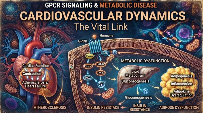
The article examines cardiovascular GPCR signaling as a dynamic response system whose abnormalities can remain hidden in resting measurements. Mechanical stretch, hormones, nutrients, sympathetic activity, and fluid balance can change the amplitude, duration, localization, and recovery of receptor signaling. Examples include mechanically activated angiotensin-receptor signaling, endosomal signaling, beta-arrestin-dependent trafficking, and biased glucagon-like peptide-one receptor responses. The article argues for dynamic challenge testing using meals, exercise, posture, glucose loads, or pharmacological stimuli, combined with timed phosphoproteomics, metabolomics, transcriptomics, imaging, and physiological measurements. Such designs can identify delayed recovery, exaggerated peaks, failed suppression, or loss of coordination across organs. Organ crosstalk among heart, kidney, pancreas, adipose tissue, and skeletal muscle is central to interpretation. Repeated interventions can test reversibility and electrical or metabolic resilience. Overall, the article frames cardiometabolic disease as a failure of signaling flexibility, timing, and recovery rather than a static abnormal concentration.
The most important characteristic of cardiovascular signaling is response to change and is hidden in a resting measurement. G protein coupled receptors are responsible for the coordination of changes in pressure, nutrients, hormones, sympathetic drive, and fluid balance; however, metabolic disease can cause changes in the rate, amplitude, and recovery of these changes before the onset of overt organ failure. The dynamics of cardiovascular signaling thus cannot be captured by simply measuring the numbers of receptors or a single biomarker in circulation. The key issue is whether stressed tissues are able to transition through signaling states efficiently with no lag time and recover to baseline once the stress has subsided.
Mechanical load is an interesting example. In addition to binding to a ligand, angiotensin II type one receptors can also be activated by mechanical stretch of cardiac tissue, and experimental studies have demonstrated that stretch is associated with beta arrestin biased signaling. Results like these make it difficult to have an easy model where receptor output is related to a hormone concentration. The signal can be influenced by pressure, wall tension, conformation of the receptor, other proteins that interact with it, and the cellular location. This can then alter the behavior of the receptors even if the circulating measurements of the ligand are the same ones that are familiar.
Add time in. Receptor activation starts at the plasma membrane; but, receptor endocytosis does not necessarily stop signalling. In several different G protein coupled receptor systems, experiments have demonstrated that signaling occurs from endosomes, resulting in two distinct stages of second messenger activity, separated in time. Trafficking and formation of receptor signaling complexes can be modified by beta arrestins, changing the timing. A short surface signal combined with an extended intracellular signal may result in a different transcriptional program than just a short pulse, and the duration is a biological variable, not a technical one.
Metabolic receptors are an example of such a process. Experimental models of work on glucagon like peptide one receptor have demonstrated that there are significant improvements to the metabolic effects of G protein biased agonists when the recruitment of beta arrestin is significantly reduced. Previously, it was found that decreased recruitment of arrestin to receptors led to decreased receptor endocytosis and to the extension of the cyclic AMP signal. These findings highlight the importance of signaling persistence and trafficking to control glucose. The receptor is not only 'on' or 'off' but can have an architecture that affects the duration of the endocrine signal.
Dynamic challenge testing is a way to bridge these mechanisms and human physiology. The response curve can be exposed by a standardized meal, exercise session, posture change, controlled glucose load, or a pharmaceutical stimulus which cannot be obtained during a fast. In addition to molecular signals, responses of insulin, glucose, catecholamines, lipids, inflammatory mediators, cardiac function and vascular responses can be tracked before, during and after challenge. There are many informative features such as, delayed recovery, exaggerated peak, failure to suppress a pathway, or loss of coordination between the organs.
When coupled at the physiological time scale, the multiomic profiling can enrich that design. Rapid kinase responses can be captured in the phosphoproteomics studies, shifts in substrates can be observed using metabolomics, later regulatory adaptation can be observed via transcriptomics while targeted proteomics can quantify receptor related signaling components. Single cell assays can determine if the dominant abnormality is in the endothelial cells, immune cells, cardiomyocytes or adipose associated cells. Then a trajectory can be obtained with repeated measures, an essential requirement when the phenomenon of interest is metabolic flexibility.
A complementary scale is given by imaging. Molecular dynamics can be linked to tissue function by echocardiographic, vascular ultrasound, magnetic resonance or metabolic imaging methods. An abnormal sign becomes clinically significant if it follows impaired myocardial relaxation, abnormal myocardial perfusion, vascular stiffness and/or ectopic lipid burden. Imaging can also help to differentiate compensation from injury. An elevated signal marker in the context of successful adaptation does not have the same meaning as it does in the context of progressive structural deterioration.
There is a need for temporal discipline of circulating biomarkers as well. Production, clearance, organ function, treatment and disease state are factors that affect the levels of natriuretic peptides, inflammatory proteins, metabolites and endocrine signals. Evaluating a single concentration can only tell if the cardiovascular system is coping well or is getting close to the stress level. When a controlled stimulus is used, prospective sampling can be conducted to determine if a biomarker can predict the transition from reversible dysfunction to persistent dysfunction. That design can relate tissue biology to clinically observable progression, instead of the correlation at one visit.
The key is organ crosstalk since cardiometabolic disease is tissue independent. There is a continuous remodeling of the adipose tissue in response to hormone and cytokine exposure, volume regulation by the kidney, renin angiotensin signaling, nutrient handling by pancreatic islets, glucose disposal by skeletal muscle and continuous response to energetic and mechanical demands by the heart. GPCR pathways span all of the compartments. A key question in a useful dynamic model is whether abnormal signaling in one organ comes before or after dysfunction in another organ, and to determine the causal order, that is, which happens first. This requires repeated observations and mechanistic experiments to further delineate causal order.
Reversibility can be directly tested in intervention studies. The value of a dynamic signalling feature as an early response marker increases if it is normalized before structural disease improves, such as through weight reduction, improved glycaemic control, exercise training, or blood pressure treatment, or through a receptor targeted therapy. Normalization is a necessary failure which can identify persistent biological risk. These studies should be prespecified as to time since a pathway can improve for a period of time and then get worse again with chronic exposure. The strongest evidence for reversibility occurs if the molecular, physiologic, and clinical processes are all in the same direction.
Dynamic measures can be transferred between studies if a standard measure is used. Response curves can be altered by challenge intensity, duration of the fast, withholding of medications, timing of the samples, the type of assay platform used, and recovery window. A protocol which only measures a peak may miss delayed normalization and a protocol which samples too late may miss rapid signaling events. Reproducible time grids and pre-specified summary features (e.g., peak amplitude, time to peak, area under the response curve and recovery slope) can allow for molecular dynamics to be compared across cohorts. An additional requirement is that the technical replicates are as important as reference materials when the phosphoproteomic or metabolomic signals are predicted to vary in minutes.
The order of events must be maintained in the Analytical Model. Both latent class and mixed effects models can be used to estimate the individual trajectory of response; and latent class models can be used to identify groups that are characterized by a different trajectory of response. Time varying mediation can be used to determine whether a molecular change comes before an imaging or physiological change or just in parallel with it. Machine learning can enhance pattern recognition, but logicality is still important as a clinically useful signature must provide information about which pathway or phase of recovery has risk. Random data splitting is not sufficient for validation—must be a separate cohort that is challenged in the same manner.
A dynamic system also helps to elucidate the concept of safety. Excessive activation of receptor pathways that promote increased acute contractility, insulin secretion and/or raised vascular relaxation will be damaging. Overshoot is easily detected in challenge studies as well as impaired response.
Heart rate, blood pressure, heart rhythm, glucose, renal markers and symptom monitoring together with molecular assays aid in discerning beneficial flexibility from an unstable physiology. The optimum signaling profile is therefore not full activation. It is a response at an appropriate scale that increases when there is a demand, synchronizes within organs and subsides when the demand subsides.
Adaptation that can be detected after a month, not minutes, can be uncovered through repeated challenge. The metabolic treatment response kinetics can be demonstrated pre and post treatment such as showing if the responses change to a new normal, stay rigid, or change to another compensatory pattern. The within-person comparison diminishes the noise from the stable, biological differences, and may also reveal early benefit before major clinical events. Correlating those changes with subsequent ones would determine if dynamic receptor phenotypes are reversible intermediate steps between metabolic stress and cardiovascular disease.
In the field of research conducted by Robert J. Lefkowitz, G protein coupled receptors are considered as regulated signals systems instead of simple switches. The difficulty with this is that the same kinds of mechanisms are observed in metabolic disease when you try to study them. Dynamic challenge tests, timed multiomics, imaging and longitudinal outcomes can reveal responses to stress that are not detectable by static profiling. The significance of the receptor behaviour comes when it is assessed as a variable event that demonstrates flexibility, threshold failure, organ crosstalk, and recovery.
CITE THIS ARTICLE
Tasmeem, S. (2026). Cardiovascular Signaling Dynamics Linking GPCR Signaling to Metabolic Disease. https://www.sadiaresearchhub.com/article/caf84733-2dc7-5e57-b26d-93d925ce7e1a
S. Tasmeem, “Cardiovascular Signaling Dynamics Linking GPCR Signaling to Metabolic Disease,” 2026. [Online]. Available: https://www.sadiaresearchhub.com/article/caf84733-2dc7-5e57-b26d-93d925ce7e1a
Tasmeem, S. (2026) ‘Cardiovascular Signaling Dynamics Linking GPCR Signaling to Metabolic Disease’. Available at: https://www.sadiaresearchhub.com/article/caf84733-2dc7-5e57-b26d-93d925ce7e1a
Tasmeem S. Cardiovascular Signaling Dynamics Linking GPCR Signaling to Metabolic Disease. 2026. https://www.sadiaresearchhub.com/article/caf84733-2dc7-5e57-b26d-93d925ce7e1a
@misc{article_caf847332dc75e57b26d93d925ce7e1a,
  title = {Cardiovascular Signaling Dynamics Linking GPCR Signaling to Metabolic Disease},
  author = {Sadia Tasmeem},
  year = {2026},
  url = {https://www.sadiaresearchhub.com/article/caf84733-2dc7-5e57-b26d-93d925ce7e1a},
}
TY  - GEN
AU  - Sadia Tasmeem
TI  - Cardiovascular Signaling Dynamics Linking GPCR Signaling to Metabolic Disease
PY  - 2026-02-22
UR  - https://www.sadiaresearchhub.com/article/caf84733-2dc7-5e57-b26d-93d925ce7e1a
ER  -
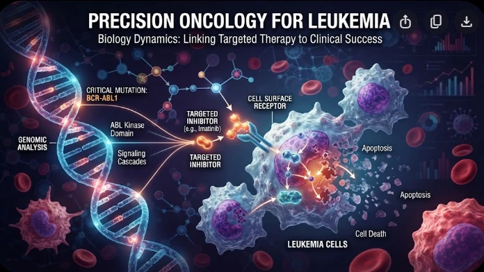
The article examines leukemia as an evolving population under targeted therapy, using chronic myeloid leukemia as the main example. BCR–ABL1 inhibition can produce deep responses, but every treatment changes the competitive environment among malignant clones. Quantitative transcript monitoring provides a continuous measure of disease burden, while deep sequencing can identify emerging resistance mechanisms before hematologic progression. A detected mutation is clinically meaningful only when its
The article examines how cardiovascular genetic risk becomes biologically active through cell-specific inflammatory signaling. Because many vascular-risk variants lie in regulatory DNA rather than coding regions, their effects may appear only in particular endothelial, macrophage, smooth-muscle, or lymphocyte states. Single-cell studies of human atherosclerotic lesions and multiomic endothelial experiments are used to illustrate this context dependence. The article also discusses IL6-related sig
The article examines steroid receptors as dynamic transcriptional systems in hormone-dependent cancers rather than simple on–off biomarkers. Estrogen, androgen, progesterone, and glucocorticoid receptors alter gene expression through ligand-induced conformation, DNA binding, chromatin accessibility, coactivators, corepressors, and signaling crosstalk. Particular attention is given to SRC-family coactivators, enhancer architecture, growth-factor pathways, protein turnover, and mutations that chan