The article links cardiac ion-channel biology with arrhythmia risk in metabolic disease. Sodium, calcium, and potassium currents must be precisely timed, and abnormalities can arise from channel mutations, accessory proteins, membrane targeting, post-translational modification, or metabolic stress. Nav1.5 is used to illustrate how reduced sodium current can slow conduction while persistent current can prolong repolarization and increase calcium load. Diabetes and obesity can modify channel behavior through oxidative stress, signaling pathways, autonomic changes, and tissue remodeling. The article recommends prospective metabolic phenotyping alongside electrocardiography, rhythm monitoring, molecular biomarkers, and chamber-specific electrophysiologic analysis. Human genetics and functional channel assays can help separate inherited susceptibility from acquired metabolic effects. The discussion also emphasizes drug safety because channel-targeting therapies can create new proarrhythmic risks. Overall, cardiometabolic arrhythmia is framed as instability in an integrated network of ion channels, membrane organization, calcium handling, metabolism, and whole-heart electrical function.
The cardiac action potential relies on precise temporal movement of sodium, calcium and potassium across the cardiomyocyte membrane. Thus, mutations in one ion channel could lead to arrhythmia, while the location of the channel, the way in which it is modified, and the effect of metabolism on the channel can also lead to arrhythmia. In addition to the stresses of cardiometabolic disease, there are multiple layers of stress, which may be why the ion channel signatures could be useful in explaining the higher incidence of abnormal rhythm in people who have diabetes and obesity.
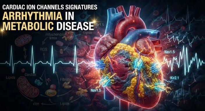
The sodium channel Nav1.5 controls the fast depolarization of the working myocardium. Decreased sodium current may lead to conduction delay and reentry, and persistent sodium current may cause a prolongation of repolarization and calcium load. The behaviour of the channels is as dependent as the pore forming gene itself on the associated proteins and membrane targeting.
Ankyrin proteins prove that is the case. Ankyrin B is involved with the organization of membrane transporters that participate in sodium and calcium homeostasis, and ankyrin G is responsible for targeting Nav1.5. Ankyrin B dysfunction may lead to sinus node dysfunction, arrhythmias of the ventricles and risk of sudden death. The mechanism is not a single current site, but rather a disruption of coordination of multiple membrane proteins.
The same machinery can be altered by the metabolic disease. Hyperglycemia leads to elevated protein O GlcNAcylation and elevation of oxidative signaling, both of which impact sodium and potassium currents. Diabetic hearts in experiment demonstrate electrical remodeling which may result in lengthening of the Action Potential duration and vulnerability to arrhythmias. This type of change has direct links to channel function and metabolic state.
The example of O GlcNAc modification of Nav1.5 is a specific example. In diabetic animal models, there is an association between the increased modification of the sodium channel and abnormal current and susceptibility to ventricular arrhythmias. The discovery demonstrates the ability of a metabolic signal to modify an electrophysiological protein without changing its DNA sequence.
Potassium channels are responsible for the repolarization and resting potential. Transient outward and inward rectifier potassium currents may be altered by hyperglycemia through CaMKII, ROS and PKC mechanisms. There can be changes in several currents which can interact and make it difficult to deduce electrocardiographic risk from one measurement of the current.
Efficient handling of calcium is also key. Calcium is taken up by L channels and increased by release from sarcoplasmic reticulum. Delayed afterdepolarizations are caused by abnormal calcium in the cell. A link between metabolic disturbance and triggered arrhythmia can be established by the increased spontaneous calcium release due to diabetes, oxidative stress, or altered CaMKII activity.
The direct connection between cellular energy and excitability is via KATP channels. These channels open in response to the ATP/ADP ratio and are opened during metabolic stress. Ankyrin B moreover affects membrane expression of KATP channels. Metabolic sensing and altered channel organization can then cause a change in rhythm in ischemia or diabetes.
Multiple layers should be incorporated into a useful ion channel signature. It can be due to genetic variants, abundance of channels, posttranslational modification, localization, electrophysiological current, and electrocardiographic phenotype. Analysis of only mRNA levels may be inadequate to detect altered trafficking, and patch clamp does not provide information about the upstream metabolic mechanism.
These layers can be linked in humans through prospective metabolic phenotyping. Monitoring glucose control, insulin resistance, lipid status, kidney function and autonomic tone are possible in addition to electrocardiograms and circulating biomarkers. The repeated monitoring can be used to determine if the deterioration of metabolic state is the cause of QT prolongation, ectopy, or other rhythm disturbances.
Single cell profiling of tissues has the potential to determine which cell populations in the heart have dysfunctional responses to stress, but sampling the heart tissue is challenging in humans. Mechanistic information can be obtained from animal models and explanted tissue, and clinical information can be obtained from circulating markers and imaging. Translation is not based on one to one assay but on concordance between these sources.
Training CMA can examine if channel related changes contribute to the relationship between metabolic disease and arrhythmia. These models must be developed with careful assumptions as the use of medication, coronary disease, autonomic dysfunction, and structural heart disease also impact on rhythm. To establish mechanism, experimental perturbation is still required.
Electrophysiological phenotyping also has the ability to distinguish conduction from repolarization defects. QRS widening is indicative of altered ventricular conduction, and QT changes are indicative of repolarization amongst others. Ambulatory recordings are able to identify pauses, atrial arrhythmias, and ventricular ectopy. A single label for arrhythmia will not mask the different mechanisms if they are linked to each phenotype.
One of the most important cardiometabolic modifiers is autonomic neuropathy. Normal heart rate variability and sympathetic parasympathetic balance may be lost in diabetes. Changes in autonomic activity affect channel phosphorylation and calcium handling. An autonomic measurement-free channel signature can identify neural effects as coming from the cardiomyocyte itself.
Yet another link between metabolism and rhythm is provided by electrolyte homeostasis. Kidney disease, diuretics and insulin treatment can affect potassium and magnesium levels, which can cause a change in membrane potential and repolarization. Large short term effects may be seen as a result of arrhythmia studies and serial electrolyte measurements should be performed.
Genotype first studies can look through rare or common variants, function. Animal models with a human ankyrin B variant can be used to investigate whether the allele modifies the rhythm in the absence of metabolic stress or when challenged with metabolic stress. These experiments are particularly helpful when predictions of the ability to cause disease are still not absolutely certain.
The measurement of cellular trafficking should be taken directly. By immunostaining and super resolution imaging, it will be determined if Nav1.5 or transporters end up in the right membrane domain. Electrophysiology and transcript/bulk proteomic data do not always agree and an abnormal total protein level, with an abnormal localization, can be responsible for this.
Then it's time for risk models to test incremental value. Mechanistic interest is not justification for a clinical assay if measurements in the channel do not improve the prediction beyond electrocardiography, imaging, metabolic markers, and clinical history. To achieve precision, there must be an additional decision value.
It is not acceptable to assume that the signature of atrial and ventricular tissue is the same. Metabolic stress may yield different responses when there is expression of potassium currents, calcium handling proteins, and structural proteins in the chamber. The sampling and modelling should then coincide with the arrhythmia phenotype.
Changes in apparent channel effects caused by fibrosis may be related to changes in electrotonic coupling between cells. An intracellular current recorded in an isolated cardiomyocyte may react differently within a fibrotic tissue. The optical mapping of intact hearts can relate the changes in cellular currents with conduction velocity and reentry at the tissue level.
Another possibility is the redox state of mitochondria. ROS affects CaMKII and channel proteins, and reduced ATP production affects pumps and KATP channels. Redox and energy markers can be measured to reveal whether electrophysiological change is along a metabolic stress pathway or if it is a simple channel defect.
Continuing collection of CGM data with rhythm data could be performed on prospective cohorts. Temporal association of glucose excursions with ectopy can lead to hypotheses about the acute triggers, while the longer-term glycemic markers reflect chronic remodeling. The two time periods are required to distinguish between trigger and substrate.
Compensatory current changes should be monitored in intervention studies. Over time, one channel may be blocked, causing other channels to remodel. The short-term antiarrhythmic effect may, therefore, differ from the long term effect. This adaptation can be identified via repeated electrophysiological measures before clinical risk levels change.
Also temperature, pH and fatty acid composition may affect the channel function and these vary in metabolic stress or ischemia. These should be controlled or modeled specially in experimental studies. Any current change due to hyperglycemia can, otherwise, be a secondary change in cellular environment. Good control enhances translation from single cardiomyocytes to whole hearts.
The final interpretation should link a molecular change with a measurable electric result, rather than just assume that altered channel expression is a marker of arrhythmia risk.
This integrated perspective is reflected in the research interests of Peter J. Mohler, whose studies are directed toward the cardiac ion channels, arrhythmia and the molecular aspects of cardiology. Not all inherited channelopathies have a channel signature. The same electrophysiological network can be modified, trafficked and signalled by metabolic stress. The validity of cardiometabolic risk is enhanced when channel biology is viewed as an integrated system among metabolism, membrane organization, calcium balance and whole heart rhythm.
Mechanism should continue to be connected with rhythm.
CITE THIS ARTICLE
Tasneem, S. (2025). Cardiac Ion Channels Signatures through Arrhythmia in Metabolic Disease. https://www.sadiaresearchhub.com/article/0d68fa1f-a248-5a1c-9155-1e261a242713
S. Tasneem, “Cardiac Ion Channels Signatures through Arrhythmia in Metabolic Disease,” 2025. [Online]. Available: https://www.sadiaresearchhub.com/article/0d68fa1f-a248-5a1c-9155-1e261a242713
Tasneem, S. (2025) ‘Cardiac Ion Channels Signatures through Arrhythmia in Metabolic Disease’. Available at: https://www.sadiaresearchhub.com/article/0d68fa1f-a248-5a1c-9155-1e261a242713
Tasneem, Sadia. “Cardiac Ion Channels Signatures through Arrhythmia in Metabolic Disease.” 2025, https://www.sadiaresearchhub.com/article/0d68fa1f-a248-5a1c-9155-1e261a242713.
Sadia Tasneem. “Cardiac Ion Channels Signatures through Arrhythmia in Metabolic Disease.” (2025). https://www.sadiaresearchhub.com/article/0d68fa1f-a248-5a1c-9155-1e261a242713.
Tasneem S. Cardiac Ion Channels Signatures through Arrhythmia in Metabolic Disease. 2025. https://www.sadiaresearchhub.com/article/0d68fa1f-a248-5a1c-9155-1e261a242713
@misc{article_0d68fa1fa2485a1c91551e261a242713,
  title = {Cardiac Ion Channels Signatures through Arrhythmia in Metabolic Disease},
  author = {Sadia Tasneem},
  year = {2025},
  url = {https://www.sadiaresearchhub.com/article/0d68fa1f-a248-5a1c-9155-1e261a242713},
}
TY  - GEN
AU  - Sadia Tasneem
TI  - Cardiac Ion Channels Signatures through Arrhythmia in Metabolic Disease
PY  - 2025-03-30
UR  - https://www.sadiaresearchhub.com/article/0d68fa1f-a248-5a1c-9155-1e261a242713
ER  -
The article examines leukemia as an evolving population under targeted therapy, using chronic myeloid leukemia as the main example. BCR–ABL1 inhibition can produce deep responses, but every treatment changes the competitive environment among malignant clones. Quantitative transcript monitoring provides a continuous measure of disease burden, while deep sequencing can identify emerging resistance mechanisms before hematologic progression. A detected mutation is clinically meaningful only when its
The article examines how cardiovascular genetic risk becomes biologically active through cell-specific inflammatory signaling. Because many vascular-risk variants lie in regulatory DNA rather than coding regions, their effects may appear only in particular endothelial, macrophage, smooth-muscle, or lymphocyte states. Single-cell studies of human atherosclerotic lesions and multiomic endothelial experiments are used to illustrate this context dependence. The article also discusses IL6-related sig
The article examines cardiovascular GPCR signaling as a dynamic response system whose abnormalities can remain hidden in resting measurements. Mechanical stretch, hormones, nutrients, sympathetic activity, and fluid balance can change the amplitude, duration, localization, and recovery of receptor signaling. Examples include mechanically activated angiotensin-receptor signaling, endosomal signaling, beta-arrestin-dependent trafficking, and biased glucagon-like peptide-one receptor responses. The