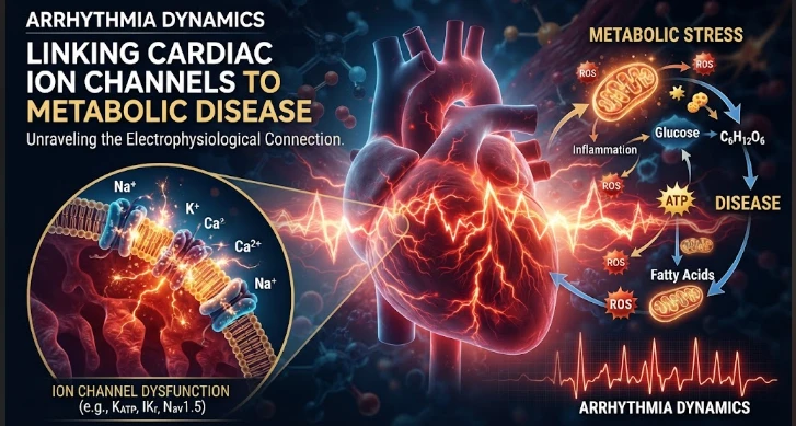
The article examines arrhythmia as a dynamic consequence of changing metabolic and electrophysiological stress. Acute hyperglycemia, oxidative signaling, autonomic shifts, electrolyte changes, and inflammation can rapidly modify ion-channel phosphorylation or gating, while chronic diabetes can alter channel expression, membrane trafficking, fibrosis, and structural substrate. Because acute and chronic mechanisms differ, the article argues for repeated measurements rather than a single electrocardiogram or metabolic value. Glucose, lipids, kidney function, electrolytes, medications, QT behavior, calcium handling, ectopy, and rhythm burden should be followed over time. Recovery after metabolic stress is treated as an important measure of electrical resilience: delayed normalization may indicate persistent remodeling. Treatment changes must be modeled carefully because glucose-lowering drugs, diuretics, beta blockers, and antiarrhythmics can alter the same pathways. The central message is that metabolic arrhythmia risk is best understood as a trajectory from reversible electrical instability to durable remodeling, with temporal order clarifying mechanism.
The risk of arrhythmia in metabolic disease is variable over time as the heart is continually adjusting to changes in glucose, lipids, oxidative, autonomic and structural stress. Rapidly: Ion channels respond to changes through phosphorylation and other modifications, and slowly through changes in expression, membrane trafficking. This may thus shift the patient from transient electrophysiologic instability to chronic electrical remodeling. Dynamic measurements are required to differentiate those stages.
Acute hyperglycemia may alter cardiac potassium currents in a short time via O GlcNAc dependent signaling, CaMKII activation, ROS, and PKC, and chronic diabetes may ultimately lead to a decrease in the expression of several components of potassium channels. Immediate and chronic effects don't necessarily go together, and thus, no single laboratory measurement can address the entire process of the disease.
Sodium current also varies with metabolic stress. Conduction and susceptibility to arrhythmia are influenced by modification of Nav1.5 during diabetes. Sustained entry of Na+ can lead to an increase in intracellular Na+ that indirectly affect Ca2+ handling via the Na+/Ca2+ exchanger. If a change occurs in one current of a chain of currents, it can propagate through the chain of currents, or along the excitation contraction network.
The dynamics of calcium molecules provides a quick pathway to arrhythmia. Delayed afterdepolarizations can be caused by increased intracellular calcium or abnormal sarcoplasmic reticulum release of calcium. CaMKII serves as a connecting link between oxidative/metabolic stress and calcium-handling proteins. Recurrent episodes can lead to remodeling which may continue for longer than the initial metabolic insult.
Ankyrin dependent targeting is a spatial component. Local ion fluxes must be coordinated between the different membrane domains of cardiomyocytes, which requires the proper localization of the ion channels and transporters. The sodium calcium exchanger, sodium potassium ATPase, inositol trisphosphate receptor and KATP channels may be disrupted by Ankyrin B dysfunction. Thus, an electrical failure may arise even though the structure of the proteins is not altered.
Dynamic challenge testing can reveal the hidden reserve. Rhythm instability that is not seen at rest may be revealed by exercise, glucose loading, controlled pacing or autonomic stimulation. The electrocardiogram may provide more useful information than a baseline ECG. An individual who has normal RIs but exaggerated RIs under stress has a different risk profile than an individual with fixed abnormalities.
Transient events can be recorded by continuous or repeated electrocardiographic (ECG) monitoring. Heart rate, ectopy, Afib, and certain interval changes are measurable with wearable devices and ambulatory monitors throughout the day. These data require clinical validation at the level of clinical grade validation, but have a time axis that matches the inherent fluctuating nature of metabolic physiology.
Upstream drivers can be looked for using multiomic measurements. Electrophysiological trajectories can be associated with metabolites, inflammatory proteins, lipid species and genetic variants. A marker that is associated with a known mechanism rather than merely related to arrhythmia is most useful. The use of molecular data along with direct electrical measurements minimises the danger of building a prediction without any biological explanation.
Imaging provides the structural context. Conduction heterogeneities can occur due to fibrosis, ventricular hypertrophy, fat infiltration and ischemic injury, apart from ion channel changes. Electrocardiographic and echocardiographic characteristics can differentiate an electrical phenotype from a structural substrate dominant phenotype. Often both occur in metabolic disease and interaction models are necessary.
Medication changes should be taken into consideration with longitudinal analysis. Several medications, including glucose lowering drugs, diuretics, beta blockers, antiarrhythmics and electrolytes can alter rhythm risk. Treatment may alter potassium and magnesium levels, kidney function or autonomic status. Those covariates, if they are not taken into account, could create a spurious temporal relationship between glucose and arrhythmia.
Experimental models can be used to directly test candidate trajectories. Patch clamp and calcium imaging can be used to track the time evolution of cardiomyocytes under controlled glucose and/or oxidative stress. Animal models can link cellular changes to whole heart structural remodeling and rhythm. Reverting the metabolic stress may reveal the changes in the electrical status that may be restored or permanent.
There is an important difference between reversible and irreversible diseases, clinically. If the patient's channel modification can be normalized by better metabolic control and the susceptibility to arrhythmia is decreased, the pathway is potentially modifiable. The critical importance of earlier intervention increases if fibrosis and channel remodeling continue despite metabolic improvement. Longitudinal studies can be used to define a point where transient stress transforms to fixed substrate.
Metabolism and electrophysiology may be affected by circadian timing. There are diurnal changes in glucose, autonomic tone, hormone levels and channel expression. Testing done at different times may add noise or false trends. Longitudinal studies may be improved by either standardizing timing or by explicit circadian modelling.
Exercise is physiologically relevant as the catecholamines, calcium cycling and energy demand are all increased in concert. LIMCA and/or calcium instability may be revealed by EIE. Recovery kinetics after exercise may offer clues to the rate of electrical system return to baseline.
Another dynamic modifier is sleep. Obesity is associated with periods of hypoxia and sympathetic surges, which are characteristic of OSA. Arrhythmia burden is independent of glucose control as sleep apnea is treated. This exposure should be recorded in metabolic arrhythmia models and not be considered as a complete explanation for body mass.
The reversibility of the condition can be tested molecularly by repeated tissue or cellular sampling. When O GlcNAcylation occurs following the return to normal glucose and potassium currents recover, the pathway is plastic. Persistent current abnormalities despite normoglycemia indicate slower remodeling/structural change.
Event based analysis can make molecular and electrical data coincide. Rather than an average for a week of glucose and an average for a week of rhythm, continuous monitoring can determine if there is ectopy preceding glucose rises within a certain window over a specific period of time. This analysis can produce hypotheses on acute triggers and follow-up has a longer time frame for chronic substrate.
Dynamic modeling can be based on state transitions like stable rhythm, ectopy, nonsustained tachycardia, and sustained arrhythmias. Metabolic and molecular features can be related to the probability of moving between states. This framework is a more natural way of reflecting progression than a single binary outcome.
Post stress is as instructive as pre stress. Once glucose is normalized or acute illness is over, the researchers can assess the speed at which QT returns to normal, the speed of calcium handling recovery and channel changes return to normal. A slow recovery may be a sign of low electrical resilience.
In animal models, autonomic blockade experiments can differentiate direct myocardial effects from sympathetic or parasympathetic mediation. Once autonomic influence is controlled, a metabolic challenge will be less likely to influence arrhythmia, unless there are intrinsic cardiac mechanisms involved.
Ordinary physiological challenges – such as food or physical activity – can be used in human challenge studies with caution. Transient repolarization changes can be recorded upon repeated ECG around these events without invasive procedures. Standardisation facilitates comparison between participants.
Clinically important events, rather than surrogate intervals, should be considered when validating a trajectory. It is hoped that a model predicting QT variability will be tested against arrhythmia burden or sudden cardiac outcome in the future. Surrogate endpoints can be beneficial mechanistically, but should not be used indefinitely.
Can benefit efficiency in long studies using adaptive sampling. Those who are deteriorating metabolically fast or new rhythm abnormalities can be monitored more closely and stable participants can be monitored less. Outcome driven bias is avoided by predefined rules. This design can observe the transition into electrical instability without having to constantly test the participants' electrical instabilities arduously.
Dynamic evidence is most compelling when molecular change, electrical instability, and clinical events occur over a comprehensible time course in repeated observation.
Peter J. Mohler's research focus on ion channel organization and arrhythmia supports this time dependent view. The progression of cardiac electrophysiology in metabolic disease should be treated as a continuum of channel modification, membrane targeting, calcium handling, autonomic input and structural change. When a system reaches its "reserve", it can be detected through dynamic challenge tests and through molecular measurements over time. Arrhythmia then is not a single unprovoked occurrence, but an observable culmination of the electrical adaptation which has been occurring in response to metabolic stress.
The path turns into the main phenotype.
Multiple measurements make causal sequence clear.
CITE THIS ARTICLE
Tasneem, S. (2025). Arrhythmia Dynamics Linking Cardiac Ion Channels to Metabolic Disease. https://www.sadiaresearchhub.com/article/819c801a-62f4-57bd-adf1-49909ec8a4fe
S. Tasneem, “Arrhythmia Dynamics Linking Cardiac Ion Channels to Metabolic Disease,” 2025. [Online]. Available: https://www.sadiaresearchhub.com/article/819c801a-62f4-57bd-adf1-49909ec8a4fe
Tasneem, S. (2025) ‘Arrhythmia Dynamics Linking Cardiac Ion Channels to Metabolic Disease’. Available at: https://www.sadiaresearchhub.com/article/819c801a-62f4-57bd-adf1-49909ec8a4fe
Tasneem, Sadia. “Arrhythmia Dynamics Linking Cardiac Ion Channels to Metabolic Disease.” 2025, https://www.sadiaresearchhub.com/article/819c801a-62f4-57bd-adf1-49909ec8a4fe.
Sadia Tasneem. “Arrhythmia Dynamics Linking Cardiac Ion Channels to Metabolic Disease.” (2025). https://www.sadiaresearchhub.com/article/819c801a-62f4-57bd-adf1-49909ec8a4fe.
Tasneem S. Arrhythmia Dynamics Linking Cardiac Ion Channels to Metabolic Disease. 2025. https://www.sadiaresearchhub.com/article/819c801a-62f4-57bd-adf1-49909ec8a4fe
@misc{article_819c801a62f457bdadf149909ec8a4fe,
  title = {Arrhythmia Dynamics Linking Cardiac Ion Channels to Metabolic Disease},
  author = {Sadia Tasneem},
  year = {2025},
  url = {https://www.sadiaresearchhub.com/article/819c801a-62f4-57bd-adf1-49909ec8a4fe},
}
TY  - GEN
AU  - Sadia Tasneem
TI  - Arrhythmia Dynamics Linking Cardiac Ion Channels to Metabolic Disease
PY  - 2025-03-23
UR  - https://www.sadiaresearchhub.com/article/819c801a-62f4-57bd-adf1-49909ec8a4fe
ER  -
The article examines leukemia as an evolving population under targeted therapy, using chronic myeloid leukemia as the main example. BCR–ABL1 inhibition can produce deep responses, but every treatment changes the competitive environment among malignant clones. Quantitative transcript monitoring provides a continuous measure of disease burden, while deep sequencing can identify emerging resistance mechanisms before hematologic progression. A detected mutation is clinically meaningful only when its
The article examines how cardiovascular genetic risk becomes biologically active through cell-specific inflammatory signaling. Because many vascular-risk variants lie in regulatory DNA rather than coding regions, their effects may appear only in particular endothelial, macrophage, smooth-muscle, or lymphocyte states. Single-cell studies of human atherosclerotic lesions and multiomic endothelial experiments are used to illustrate this context dependence. The article also discusses IL6-related sig
The article examines cardiovascular GPCR signaling as a dynamic response system whose abnormalities can remain hidden in resting measurements. Mechanical stretch, hormones, nutrients, sympathetic activity, and fluid balance can change the amplitude, duration, localization, and recovery of receptor signaling. Examples include mechanically activated angiotensin-receptor signaling, endosomal signaling, beta-arrestin-dependent trafficking, and biased glucagon-like peptide-one receptor responses. The