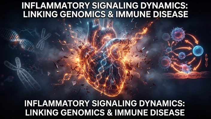
The article examines how cardiovascular genetic risk becomes biologically active through cell-specific inflammatory signaling. Because many vascular-risk variants lie in regulatory DNA rather than coding regions, their effects may appear only in particular endothelial, macrophage, smooth-muscle, or lymphocyte states. Single-cell studies of human atherosclerotic lesions and multiomic endothelial experiments are used to illustrate this context dependence. The article also discusses IL6-related signaling, clonal hematopoiesis, inherited modifiers, and spatial localization within plaque architecture. Functional perturbation with CRISPR, receptor blockade, gene silencing, and human vascular models is proposed to test whether nominated regulators actually alter adhesion, migration, cytokine release, barrier function, or plaque-relevant behavior. Timing, vascular-bed differences, ancestry, dose-response relationships, and uncertainty in causal inference require explicit treatment. The overall framework seeks a directed path from genetic variant to regulatory state, cell behavior, tissue change, and clinical outcome rather than relying on generalized inflammatory associations.
Selected Category: C
Blood-based markers of the inflammatory process like C reactive protein or interleukin 6 are frequently used to summarize inflammatory signaling in vascular disease, but do not provide information about the cell that received the signal, the program of regulation that was altered or the vascular compartment that was vulnerable. The genetic component introduces another layer of complexity as the effects of genetic variation can be cell specific. A variant that alters the expression of a gene may affect only monocytes but not endothelial cells, or a variant may be effective only once inflammatory stimulation has been given. To achieve precision, the genotype must be mapped to the cell state, not the average of the whole blood.
GWAS studies have been used to identify numerous genetic variants associated with CAD and stroke and related traits, but the majority of these variants are not within coding regions. They are frequently regulated by regulatory DNA, binding sites of transcription factors and chromatin accessibility. A risk variant may only affect one cell type under certain circumstances. Combining cardiovascular genetics with single cell expression and epigenomic data can then refine the statistical loci to narrow down to potential mechanisms in the macrophages, smooth muscle cells, endothelial cells or lymphocytes.
Diversity is demonstrated in human plaque studies. Multiple states of macrophages within the atherosclerotic lesions of the carotid arteries have been identified, such as inflammatory subsets, C1Q macrophages, TREM2-positive foam cells, and proliferative or smooth muscle-like programs. Biology is crude because of this heterogeneity, such that a label like the presence of macrophages is itself a biological label. When all myeloid cells are pooled it may be difficult to detect genetic effects on particular macrophage states because these states may be diluted when the proportion of each state in the population at particular disease stages is altered.
Endothelia cells are also heterogenous. We have performed multiomic experiments in primary human aortic endothelial cells under conditions that activate the cells for atherosclerosis and have uncovered distinct chromatin and transcriptomic state. Selected endothelial phenotypes were enriched for disease associated variants of coronary artery disease, which were accessible to chromatin modification. The outcome is consistent with a model of context-dependency: inheritance of a risk may become functional if an inflammatory or vascular stress event opens up a regulatory region in the cell type that can recognize the variant.
Inflammation can also be broken down genetically. The genetic proxy of lower receptor signaling of IL6 identified in the proteomic integration revealed a large protein signature in the circulation that was associated with the vascular effects, and suggested CXCL10 as a downstream mediator of IL6. These designs are not based on the concept of individual cytokine/individual outcome. A signaling pathway can leave a coordinated molecular footprint and genetic instruments can help to identify which downstream proteins are closer to the causal pathway.
The concept of cell specific modifier analyses is introduced in a second step. Clonal hematopoiesis and cardiovascular disease studies have defined inflammatory genes that inherited variants of influence the risk of specific somatic mutations. The two genes that were identified in human data as modifiers, AIM2 and IL1RAP, were consistent with the hypothesis that very same genomic lesion can cause divergent vascular outcomes across inflammatory pathway contexts. The resultant cardiovascular risk is then influenced by interactions between inherited variants, acquired mutations, and cell state.
Those interactions are determined by spatial location and will not reach vascular tissue. The blood monocyte has to attach, penetrate, transform and interact with lipids, the dead cells, the body's smooth muscle and other immune cells. The changes in gene expression occur at each transition. The spatial transcriptomics and multiplex imaging can locate inflammatory states in plaque architecture, identifying cells in the vicinity of necrotic core, fibrous cap, adventitial zone and microvessels. A pathway related to plaque rupture needs to be targeted to the tissue context in which rupture biology occurs.
Cell atlases are descriptive, and perturbation is needed. Candidates to regulate these cells can be tested in primary cells or in relevant models using CRISPR editing, cytokine blockade, receptor antagonism, or targeted gene silencing. Mechanistic weight is given to an endothelial risk locus if editing of the risk-enhancer alters the inflammatory adhesion or barrier function. The case for a myeloid pathway is strengthened if perturbation effects on cytokine release, migration, or foam cell behavior are observed. Functional rescue can help identify causal regulators from markers that are merely a by-product of disease.
Perturbation studies should be timed as well. Activation of endothelial cells can be very rapid and occurs after exposure to cytokines, whereas chromatin remodeling and phenotypic transition takes a longer time. Lipid loading and hypoxia can alter macrophage states. One single endpoint may lead to a wrong classification of adaptive response as a stable disease program. Time course experiments can reveal transient regulators, continuous pathways, and late changes that occur as a result of irreversible tissue damage.
Human tissue models provide a link between isolated cells and full plaques. Interfaces between endothelium, smooth muscle and immune cells may be maintained in engineered vessel systems, vascular organoids, plaque derived cultures or co culture systems. Genetically defined perturbations can be then subjected to studies under flow, lipid exposure or inflammatory stimulation. Such systems are not capable of recreating all the characteristics of atherosclerosis, but they allow for controlled studies of cell-specific mechanisms to be performed, which would not be possible otherwise through bulk cohort data.
Care needs to be taken at the design of the biomarker in the clinical translation. Only after a link between the presence of a plasma inflammatory protein and a given state of a vascular cell is known can a protein be used as an accessible proxy. However, if it is the same protein, then its specificity would be limited in case of infection, adipose inflammation, kidney dysfunction and plaque activity. Genotyping and cell state markers, in addition to protein panels, can help with interpretation. Validation should determine if the molecular signature adds value to traditional risk factors and independent of vascular bed in predicting progression.
Coronary, carotid and aortic, and peripheral arteries have different flow patterns, mechanical stress, and local cellular environment, which is important. No single gene regulatory program can be simply translated to a coronary disease. Conserved inflammatory pathways and bed specific mechanisms can be identified by comparing single cell and genomic studies. This distinction is significant for therapeutic targeting since a pathway that has a helpful effect in one territory might have different consequences in another.
The need for population diversity is also crucial. Variants in regulation occur at different rates, inflammatory exposures vary between communities and distributions of risk factors affect vascular state. Mechanisms from one ancestry may not be covered by the cell specific expression quantitative trait loci derived primarily from that same ancestry. Fine mapping by diverse recruitment and ancestry can enhance causal localization and minimize inequality related to genomic interpretation. A precision framework should not be based on a small reference population.
The analytical target is the central theme: a directed network from variant to regulatory state, cell behaviour, tissue change, clinical outcome. Assessments of associations should be done via a genetic approach, assays of cells should be done via an assay of cells, spatial profiling should be done via a spatial approach, perturbation should be done via a perturbation approach, and longitudinal follow up should be done via a longitudinal approach, depending on the size and type of link. When there is consensus across those layers, it's more convincing than any one ominic signal.
The intensity of a stimulus also may affect its regulatory effects. Lowly IL, interferon, lipid and/or oxidative stress will make a little variant influential. Such thresholds can be mapped with experimental dose response designs and tested for in genotype stratified human cohorts to see if similar interactions are seen in acute inflammation or chronic vascular disease. The logic of this gene by environment is explained by why the inherited cardiovascular risk may be silent for years, and only become biologically visible at a later age.
Uncertainty should be kept throughout the process of data integration. A genetic signal that is colocated can nominate a gene but does not necessarily identify the responsible cell, and an association of a single cell may identify a state but does not necessarily provide direction. Uncertainty can be propagated instead of making each layer a binary decision in a Bayesian or probabilistic network model. When multiple independent lines of evidence support a variant in a cell type, pathway, and vascular phenotype, prioritization increases.
Timothy A. McCaffrey's research interests are centered on the intersection of cardiovascular genomics and inflammation and are now increasingly being defined by the cell. When all of these elements are identified, inflammatory signaling becomes clinically relevant when the cell, the mechanism, the tissue niche and the stage of the disease are identified. The combination of genetics and perturbation based immune mapping and spatial vascular biology could reignite the transformation from generalized inflammatory associations to specific mechanisms, uncovering targets that represent actual mechanisms of vascular disease development within the heterogeneous human tissue.
CITE THIS ARTICLE
Tasmeem, S. (2026). Inflammatory Signaling Dynamics Linking Cardiovascular Genomics to Immune Disease. https://www.sadiaresearchhub.com/article/91c8cdaf-79c1-5993-9ccb-950a3c39c65f
S. Tasmeem, “Inflammatory Signaling Dynamics Linking Cardiovascular Genomics to Immune Disease,” 2026. [Online]. Available: https://www.sadiaresearchhub.com/article/91c8cdaf-79c1-5993-9ccb-950a3c39c65f
Tasmeem, S. (2026) ‘Inflammatory Signaling Dynamics Linking Cardiovascular Genomics to Immune Disease’. Available at: https://www.sadiaresearchhub.com/article/91c8cdaf-79c1-5993-9ccb-950a3c39c65f
Tasmeem S. Inflammatory Signaling Dynamics Linking Cardiovascular Genomics to Immune Disease. 2026. https://www.sadiaresearchhub.com/article/91c8cdaf-79c1-5993-9ccb-950a3c39c65f
@misc{article_91c8cdaf79c159939ccb950a3c39c65f,
  title = {Inflammatory Signaling Dynamics Linking Cardiovascular Genomics to Immune Disease},
  author = {Sadia Tasmeem},
  year = {2026},
  url = {https://www.sadiaresearchhub.com/article/91c8cdaf-79c1-5993-9ccb-950a3c39c65f},
}
TY  - GEN
AU  - Sadia Tasmeem
TI  - Inflammatory Signaling Dynamics Linking Cardiovascular Genomics to Immune Disease
PY  - 2026-01-11
UR  - https://www.sadiaresearchhub.com/article/91c8cdaf-79c1-5993-9ccb-950a3c39c65f
ER  -
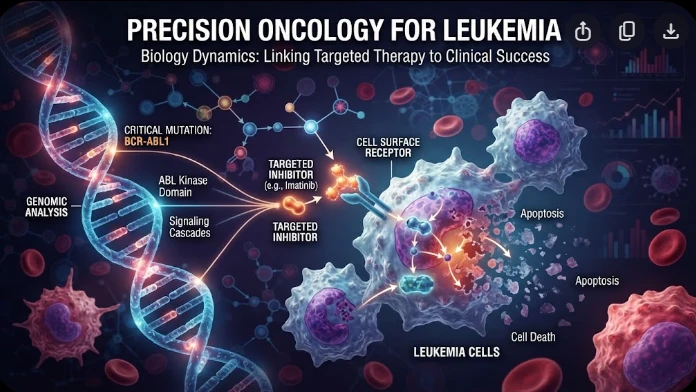
The article examines leukemia as an evolving population under targeted therapy, using chronic myeloid leukemia as the main example. BCR–ABL1 inhibition can produce deep responses, but every treatment changes the competitive environment among malignant clones. Quantitative transcript monitoring provides a continuous measure of disease burden, while deep sequencing can identify emerging resistance mechanisms before hematologic progression. A detected mutation is clinically meaningful only when its
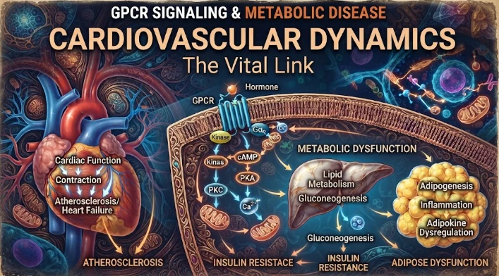
The article examines cardiovascular GPCR signaling as a dynamic response system whose abnormalities can remain hidden in resting measurements. Mechanical stretch, hormones, nutrients, sympathetic activity, and fluid balance can change the amplitude, duration, localization, and recovery of receptor signaling. Examples include mechanically activated angiotensin-receptor signaling, endosomal signaling, beta-arrestin-dependent trafficking, and biased glucagon-like peptide-one receptor responses. The
The article examines steroid receptors as dynamic transcriptional systems in hormone-dependent cancers rather than simple on–off biomarkers. Estrogen, androgen, progesterone, and glucocorticoid receptors alter gene expression through ligand-induced conformation, DNA binding, chromatin accessibility, coactivators, corepressors, and signaling crosstalk. Particular attention is given to SRC-family coactivators, enhancer architecture, growth-factor pathways, protein turnover, and mutations that chan