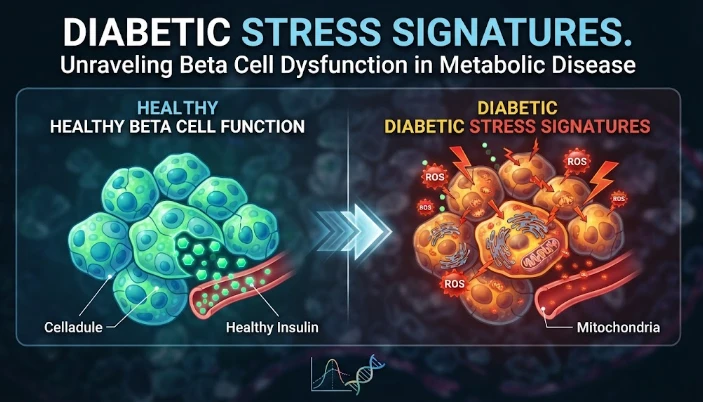
The article presents diabetic beta-cell stress as a transition from adaptive compensation to dysfunction rather than a simple consequence of high glucose. Beta cells must continually fold and process proinsulin, coordinate calcium and mitochondrial activity, and preserve mature identity under rising metabolic demand. Endoplasmic-reticulum stress and the unfolded-protein response are discussed as initially protective mechanisms that can become maladaptive when stress persists. Single-cell studies show heterogeneous beta-cell states, including potentially reversible stress adaptation before irreversible failure. The article proposes combining molecular stress signatures with glucose challenges, C-peptide, proinsulin, insulin sensitivity, continuous glucose monitoring, and other functional measures to identify reserve. Circulating biomarkers require validation against human islet biology. Experimental stressors, gene perturbation, organoid or islet models, and washout studies can test causality and reversibility. Clinically, a useful signature should predict future secretory decline and identify a stage when reducing metabolic demand can still preserve beta-cell function.
The cells of the pancreas have a tough job. Every cell needs to detect the nutrients, produce a lot of insulin, correctly fold proinsulin, package the insulin into granules, and send it out within minutes of the nutrients' arrival. Metabolic disease increases that demand. Cells can be in a state of stress that alters secretion, cell identity, protein handling, and cell survival before beta cell mass drops significantly. A diabetic stress signature should thus reflect adaptation and incipient failure and not simply a high glucose concentration after failure occurs.
The endoplasmic reticulum is a major stress sensor, since proinsulin folding takes place within it. The unfolded protein response is a cellular stress response that, at first, defends cells by limiting translation, increasing folding capacity and removing misfolded proteins only to lead to cell death in the event of a sustained increase in insulin demand. An individual or chronic stress can force the same system to become dysregulated and cause cell death. The biological issue is threshold: if there is adaptive signaling it allows for a period of function, but if it goes unresolved over a longer period of time, it becomes maladaptive.
Experimental studies of chronic reversible endoplasmic reticulum stress have demonstrated that beta cells can go through a process of reprogramming transcription and translation, lose expression of mature functional markers, and become “plastic” without completely losing their ability to recover after stress is removed. The investigators outlined a "stress adaptation exhaustion" process, and single cell analysis of islets from human type one diabetes showed that these programs of stress adaptation were disrupted. This evidence is in line with a trajectory model instead of a healthy/failed classification.
Stress signature should then contain stress markers for burden and reserve. These can include endoplasmic reticulum (ER) chaperones, unfolded protein response genes, markers of oxidative stress, insulin processing, mitochondrial function, calcium handling, and mature beta cell identity. Failure for any given type of diabetes is unlikely to be determined by just one marker. The informative pattern might be an increase in stress, loss of identity and absence of stimulus secretion.
Single cell profiling is necessary as the islets are composed of beta, alpha, delta, ductal, endothelial, immune and other cells. There may be variations in the response to stress among those populations. The increase in a bulk islet signal may be due to changes in cell state or changes in the number of immune or ductal cells. Attributed measurements, obtained at the level of the cell, retain the attribution and can identify subgroups of beta cells that are resilient, even as other groups decompensate.
Those states can be correlated with physiology by means of prospective metabolic phenotypings. Complementary information is available from glucose tolerance testing, mixed meal stimulation, continuous glucose monitoring, fasting insulin, C peptide, proinsulin and insulin sensitivity measures. The molecular stress state may occur before the first phase decline or deteriorating glycaemic variability can be seen with repeated sampling. The order of the events is important because the high glucose may exacerbate the cell's stress.
Tissue biology may become clinically available by circulating biomarkers. Islet state can be assessed using proinsulin to insulin relationships, beta cell derived proteins, extracellular vesicle cargo, and stress associated molecules. These must be validated directly against human islet biology. A marker that is not specific to the pancreas, but is correlated with the severity of the disease, may not be specific to the liver or muscle cells, or immune cells.
Causal mediation can be used to assess if stress exists between metabolic pressure and beta cell dysfunction. In obesity, or insulin resistance, the need for secretion may go up, leading to greater protein folding stress and loss of beta cell reserve. Longitudinal models can estimate if the decrease in secretion later is because of a molecular stress signature. While such analysis will not provide evidence of mechanism, it can guide pathways for intervention experiments.
Functional perturbation takes that next step. Glucose, lipids, cytokines, or endoplasmic reticulum stressors can be added to human islets, stem cell derived islets and beta cell models. Genetic modification may enhance or disrupt a candidate pathway to protect a candidate. A signature becomes mechanistic if it is able to predict dysfunction and when pathway manipulation affects survival or insulin secretion in the right direction.
Try out the reversibility explicitly. A cell state that recovers towards mature beta cell function when the stress is reduced, is no longer a cell state in which function is normal. It can be defined in vitro by washout experiments and can be found in humans through clinical interventions that lower metabolic demand. The recovery of these stress markers can be followed after weight loss, improved insulin sensitivity or treatment that reduces glucose exposure to determine if secretion improves before the markers do.
There will be considerable variation among individuals. Stress thresholds can be altered by genetic risk, age, obesity history, autoimmunity, medications and previous glycaemic exposure. Two people with the same glucose level can have varying amounts of beta cell reserve. Precision phenotyping should, therefore, target subtypes with high stress and preserved secretion, high stress and declining identity, or low measurable stress and reduced beta cell mass.
The type 1 and type 2 diabetes difference also counts. Although the inflammatory attack is a key feature of type one diabetes, and insulin resistance and nutrient stress feature prominently in many type two trajectories, both of these processes can converge on endoplasmic reticulum stress and loss of beta cell identity. Shared modules of stress may also be present in conjunction with other disease-specific upstream causes. A good signature ought to keep both shared final paths and initial context.
Intra-islet spatial organisation has a potential to shape resilience. There are electrical and paracrine signals between beta cells, and nutrients and oxygen supply is influenced by vascular proximity. A stressed cell can cause changes to the behavior of neighboring cells as well. Imaging and spatial transcriptomics can be used to test if there are specific regions of the islets, or near immune infiltration, fibrosis or altered vasculature, where dysfunctional states cluster. This context can help provide insight on why isolated cell assays may not reflect all aspects of human disease.
When clinically validating, you should be looking for prediction and not association. A stress signature obtained on a given day should predict future deterioration of secretion, escalation to therapy or transition from high risk metabolic state to diabetes. But separate cohorts should use the same assay and thresholds that were prespecified. Before the complex molecular testing can become clinically useful, it is necessary to establish superiority over existing tests (such as glucose, glycated hemoglobin, and C peptide).
A particularly early signal can be given by the protein processing. Stress can lead to a disruption of the pathway from proinsulin to mature insulin in the secretory granules, before insulin production really starts falling. Therefore, an increase in proinsulin relative to mature insulin may be due to increased demand for insulin secretion or to inefficient processing. To interpret this measure, insulin sensitivity information is needed, since the same amount of peripheral resistance may be compensated for by a greater release of proinsulin without the same amount of actual injury to the cells.
State transitions in cells should be correlated with real-life survival. Transcriptionally, a stress program can appear very tough, but actually be beneficial to the cell by slowing the translation rate and triggering repair. If reserve is already depleted, another cell that has less transcription change may be closer to apoptosis. State markers and viability in combination with longitudinal function and recovery from the removal of the stress does not allow assuming that better molecular activation equals worse disease.
Metabolic stress is also a source of memory that can happen over and over. After prolonged exposure to glucose or inflammation, beta cells may be altered such that they remain forever altered when the initial stimulus is removed. Pre-stress, adaptation and recovery chromatin profiling can determine if regulatory regions return to normal. It is possible that chronic changes in accessibility may account for the fact that some effects of poor metabolic control last beyond the duration of exposure.
Raghu G. Mirmira's work in diabetes and pancreatic beta cell biology is based on a model of stress adaptation in disease progression. Single cell profiling can determine states, a dynamic metabolic test can determine reserve, and a longitudinal biomarker can link the two. The most helpful diabetic stress signature would be a reversible transition before the irreversible loss of beta cell function, providing a window of opportunity to alter the course through a decrease in demand or proteostasis.
CITE THIS ARTICLE
Tasmeem, S. (2025). Diabetic Stress Signatures through Beta Cell Function in Metabolic Disease. https://www.sadiaresearchhub.com/article/c05cc3a9-4175-5e51-8907-b5b373b13ee2
S. Tasmeem, “Diabetic Stress Signatures through Beta Cell Function in Metabolic Disease,” 2025. [Online]. Available: https://www.sadiaresearchhub.com/article/c05cc3a9-4175-5e51-8907-b5b373b13ee2
Tasmeem, S. (2025) ‘Diabetic Stress Signatures through Beta Cell Function in Metabolic Disease’. Available at: https://www.sadiaresearchhub.com/article/c05cc3a9-4175-5e51-8907-b5b373b13ee2
Tasmeem, Sadia. “Diabetic Stress Signatures through Beta Cell Function in Metabolic Disease.” 2025, https://www.sadiaresearchhub.com/article/c05cc3a9-4175-5e51-8907-b5b373b13ee2.
Sadia Tasmeem. “Diabetic Stress Signatures through Beta Cell Function in Metabolic Disease.” (2025). https://www.sadiaresearchhub.com/article/c05cc3a9-4175-5e51-8907-b5b373b13ee2.
Tasmeem S. Diabetic Stress Signatures through Beta Cell Function in Metabolic Disease. 2025. https://www.sadiaresearchhub.com/article/c05cc3a9-4175-5e51-8907-b5b373b13ee2
@misc{article_c05cc3a941755e518907b5b373b13ee2,
  title = {Diabetic Stress Signatures through Beta Cell Function in Metabolic Disease},
  author = {Sadia Tasmeem},
  year = {2025},
  url = {https://www.sadiaresearchhub.com/article/c05cc3a9-4175-5e51-8907-b5b373b13ee2},
}
TY  - GEN
AU  - Sadia Tasmeem
TI  - Diabetic Stress Signatures through Beta Cell Function in Metabolic Disease
PY  - 2025-10-26
UR  - https://www.sadiaresearchhub.com/article/c05cc3a9-4175-5e51-8907-b5b373b13ee2
ER  -
The article examines leukemia as an evolving population under targeted therapy, using chronic myeloid leukemia as the main example. BCR–ABL1 inhibition can produce deep responses, but every treatment changes the competitive environment among malignant clones. Quantitative transcript monitoring provides a continuous measure of disease burden, while deep sequencing can identify emerging resistance mechanisms before hematologic progression. A detected mutation is clinically meaningful only when its
The article examines how cardiovascular genetic risk becomes biologically active through cell-specific inflammatory signaling. Because many vascular-risk variants lie in regulatory DNA rather than coding regions, their effects may appear only in particular endothelial, macrophage, smooth-muscle, or lymphocyte states. Single-cell studies of human atherosclerotic lesions and multiomic endothelial experiments are used to illustrate this context dependence. The article also discusses IL6-related sig
The article examines cardiovascular GPCR signaling as a dynamic response system whose abnormalities can remain hidden in resting measurements. Mechanical stretch, hormones, nutrients, sympathetic activity, and fluid balance can change the amplitude, duration, localization, and recovery of receptor signaling. Examples include mechanically activated angiotensin-receptor signaling, endosomal signaling, beta-arrestin-dependent trafficking, and biased glucagon-like peptide-one receptor responses. The