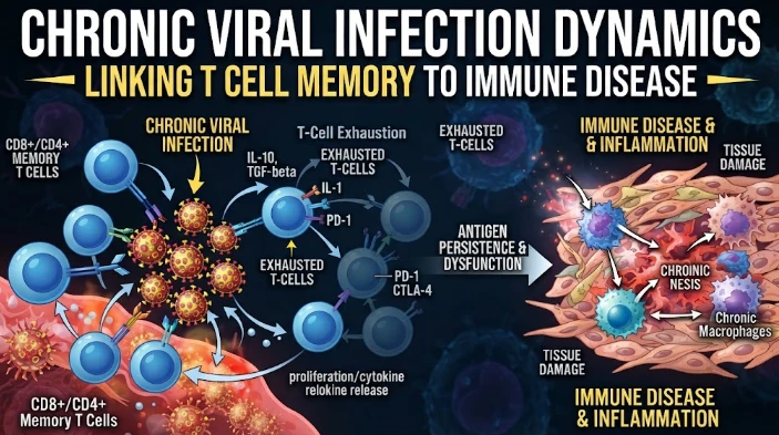
The article examines chronic viral infection through the spatial organization and differentiation of T-cell memory. It describes intermediate and exhausted T-cell populations with different proliferative capacity, tissue access, and responsiveness, emphasizing that blood measurements can miss important states in viral reservoirs and tissue niches. Persistent antigen creates hierarchies ranging from progenitor-like cells to terminally exhausted populations, with implications for checkpoint-based interventions. The article recommends combining single-cell transcriptomics, chromatin profiling, T-cell receptor sequencing, antigen localization, and spatial mapping to reconstruct cell-state trajectories. Human tissue models and perturbation experiments can test whether chemokines, antigen presentation, metabolic stress, or checkpoint pathways causally shape those states. Sampling method, tissue site, selection bias, and population heterogeneity require careful control. The central argument is that chronic infection should be understood as an immune geography in which cell state, clonotype, local antigen burden, and tissue neighborhood jointly determine whether memory populations can maintain control.
Immune geography is developed with chronic viral infection. The concentration of antigens, inflammatory signals, stromal structure, vascular access, metabolic conditions, and neighboring cells vary in the different tissues, such as blood, lymphoid tissue, liver, mucosal surfaces, and sites of viral persistence. T cell memory, then, is not stored in one population of cells, but spread throughout locations. A blood profile can provide valuable information, but it can also obscure very important cell states, and average them out, leaving out where the immune system succeeds and where the virus persists to cause dysfunction.
There are subpopulations of memory CD8 T cells with various trafficking properties and memory potential. Persistent viral infections showed that in chronic infection there was an intermediate peripheral memory population defined by the presence of CX3CR1, and that cells with similar characteristics were seen during human cytomegalovirus infection and vaccination. These populations reveal that chronic antigen exposure is not enough to generate a single exhausted compartment. There are cells that retain proliferative and effector characteristics in a space between the central memory and the terminal effector.
Receptor and transcriptional markers are read in the context of the local location, which is why location is a factor in meaning. In fact, a chemokine receptor associated with circulation can be used to mark access to vascular niches, and tissue residency markers can be used to mark long-term positioning within organs. Different compartments can be adapted to the same transcription factor, and yet be dysfunctional in the other. When efficacy relies on delivery to an infected niche as opposed to simply increasing cell numbers in circulation, spatial profiling is essential.
A hierarchy of exhausted T cell states can be produced by persistent antigen. Progenitors are capable of proliferation and proliferate to subtypes that are terminally exhausted, and terminal states have more effector characteristics and less proliferative capacity. This hierarchy is important in the case of immunotherapy, since checkpoint blockade is most effective when responsive precursor cells are still present. It is impossible to distinguish the presence of progenitors that can regenerate in a tissue from terminally dysfunctional cells or a mixture of cells that have very different therapeutic potential, from a bulk measurement of an inhibitory receptor.
Perturbation based immune mapping can overcome this issue. By studying the functional response of these cells in vitro following stimulation with specific antigen, cytokines or pathway inhibitors, changes in cell state associated with changes in function can be identified. Sequencing of the T cell receptor could link precursor and descendant populations. Single cell transcriptomics can reveal the regulatory programs that exist, and chromatin profiling can reveal whether the state is poised for recovery or whether there are stable epigenetic remodeling that constrain them. These programs are then localized in a spatial manner next to infected cells, macrophages, dendritic cells, stromal elements or fibrotic regions.
Mechanistic validation can be achieved with human tissue models, without the assumption of circulating cells to reproduce tissue biology. Selected features of cellular interaction can be maintained in organoid systems, explants, precision cut tissue preparations, or co culture models. These allow for precise regulation of chemokines, antigen presentation, metabolic stress or checkpoint pathways. A perturbation-induced change in T cell positioning or function in a model including relevant tissue components is a candidate mechanism that strengthens the credibility of the model.
The mapping of the viral load must be performed at the same spatial and temporal scale. Tissue reservoirs may continue to exist and plasma measures may decline. However, don't assume that low level nucleic detection is indicative of productive infection. Viral RNA or protein localization combined with state of immune cells can distinguish between sites of ongoing stimulation and sites with residual material. If it is possible to sample repeatedly it is possible to determine if a T cell state comes before viral control or comes after the antigen has fallen.
This concept of therapeutic response can then be viewed as traversing a state space. Antiviral therapy can help lower antigen levels; this allows for immune recovery to begin. Vaccination can allow for the expansion of selected clones or the recruitment of new clones. Responsive cells can have altered growth and effector function when the pathway is perturbed at the checkpoint. The key take-home message is not just an increase in cell numbers following treatment, but a shift toward a coordinated state that is linked to sustained control, appropriate tissue access and retained recall.
Even in a tissue focused design, the population diversity is important. There are factors that may affect cell state distributions, such as age, sex, viral history, HLA genotype, coinfection, prior treatment, and metabolic disease. A tissue signature may seem universal when it only captures one clinical subpopulation due to the sampling of just one of these subpopulations. A method of external validation should, therefore, maintain spatial definitions and assay methods. The meaning of a signature in liver tissue does not imply the meaning in blood or airway tissue; it needs to be tested.
Spatial view also translates the biomarker strategy. If markers are associated prospectively with tissue states, then they can be of value. When a good relationship is established, soluble chemokines, clonotype frequencies or transcripts in the blood could be used as accessible proxies. This connection should be examined – it can't be taken for granted. The aim is to develop a biomarker that indicates the biology of a compartment whose accessibility is limited and of known uncertainty.
Special care must be taken to tissue resident memory cells as they are not resting but rather an active program. Tissues of low re-entry of cells can be maintained by local cytokines, epithelial signals, antigen presentation and tissue metabolism. These populations can, at least in immediate terms, offer a protection at barrier sites but they can be also stimulated by persistent antigen. Matched samples of blood and tissue can be used to determine shared and compartment restricted features. A blood biomarker is more useful if it has been shown to directly correlate to the function of a tissue resident rather than by relying on a correlation with a marker.
Another form of spatial technology is the identification of neighbourhoods at the cellular level. Local cells in the vicinity of infected cells, antigen presenting cells and inflammatory macrophages are not finely regulated by the same signals as phenotypically similar cells that are fibotically separated. These neighborhoods can be quantified by multiplex imaging and spatial transcriptomics, and interactions can be computationally nominated for perturbation via ligand receptor analysis. There is still a need for the candidate interaction to be tested in the lab in order to determine that physical proximity is sufficient to indicate functional communication. Spatial association is only mechanistic evidence when the disruption of the proposed signal alters the behaviour of the cells in the expected direction.
The conclusions can be a result of sampling strategy. The different tissue depths and cell composition are represented by fine needle aspirates, biopsies, resection material, mucosal brushes and blood. The severity of the disease may also affect who will be sampled invasively, introducing selection bias.
The careful reporting of tissue acquisition, anatomical site, processing time, and cell recovery of studies are needed. Spatial state replication assists in the ability to prevent a proposed spatial state from being created by one procedure. Although it is difficult to obtain longitudinal tissue samples, it is also possible to obtain smaller paired samples to support interpretation from larger blood cohorts.
It is also possible that some apparent contradiction between studies is due to the kind of tissue that was analyzed. One group has an organ with macrophages that also has more severe T cell dysfunction, while another has an organ that also has macrophages, but which has more effective control. This difference may not be caused by analytical error, but by the state of the macrophages, the density of the antigens, the organization of the stromal, or treatment history. Such disagreements can be translated into testable hypotheses about niche specific regulation when harmonized spatial annotation and functional perturbation are combined.
State of the T cell should be used in conjunction with local antigen presentation. Dendritic cells, Macrophages and infected target cells may vary in terms of peptide display and costimulation. The problem in the population of lymphocytes may not be a failure of the intrinsic recall machinery but rather a failure in antigen presentation. The mechanisms can be distinguished by paired cellular assays.
Chronic viral infection is dynamic: Immune cells migrate, differentiate, encounter antigen and adapt to local stress. In this field, Rafi Ahmed' research has helped to establish T cell memory as a spectrum, defined not just by a single stable endpoint, but by antigen persistence and differentiation. Spatial immune mapping applies the same concept by also incorporating the location of the lineages and functions. When these aspects of cell state, tissue niche, antigen burden and therapeutic perturbation are all measured simultaneously, cell state of memory populations that actually provide control in compartments in which chronic infection exists becomes more credible.
CITE THIS ARTICLE
Tasmeem, S. (2026). Chronic Viral Infection Dynamics Linking T Cell Memory to Immune Disease. https://www.sadiaresearchhub.com/article/6a23ff7a-1041-5f84-90ce-e569784a33ad
S. Tasmeem, “Chronic Viral Infection Dynamics Linking T Cell Memory to Immune Disease,” 2026. [Online]. Available: https://www.sadiaresearchhub.com/article/6a23ff7a-1041-5f84-90ce-e569784a33ad
Tasmeem, S. (2026) ‘Chronic Viral Infection Dynamics Linking T Cell Memory to Immune Disease’. Available at: https://www.sadiaresearchhub.com/article/6a23ff7a-1041-5f84-90ce-e569784a33ad
Tasmeem, Sadia. “Chronic Viral Infection Dynamics Linking T Cell Memory to Immune Disease.” 2026, https://www.sadiaresearchhub.com/article/6a23ff7a-1041-5f84-90ce-e569784a33ad.
Sadia Tasmeem. “Chronic Viral Infection Dynamics Linking T Cell Memory to Immune Disease.” (2026). https://www.sadiaresearchhub.com/article/6a23ff7a-1041-5f84-90ce-e569784a33ad.
Tasmeem S. Chronic Viral Infection Dynamics Linking T Cell Memory to Immune Disease. 2026. https://www.sadiaresearchhub.com/article/6a23ff7a-1041-5f84-90ce-e569784a33ad
@misc{article_6a23ff7a10415f8490cee569784a33ad,
  title = {Chronic Viral Infection Dynamics Linking T Cell Memory to Immune Disease},
  author = {Sadia Tasmeem},
  year = {2026},
  url = {https://www.sadiaresearchhub.com/article/6a23ff7a-1041-5f84-90ce-e569784a33ad},
}
TY  - GEN
AU  - Sadia Tasmeem
TI  - Chronic Viral Infection Dynamics Linking T Cell Memory to Immune Disease
PY  - 2026-02-01
UR  - https://www.sadiaresearchhub.com/article/6a23ff7a-1041-5f84-90ce-e569784a33ad
ER  -
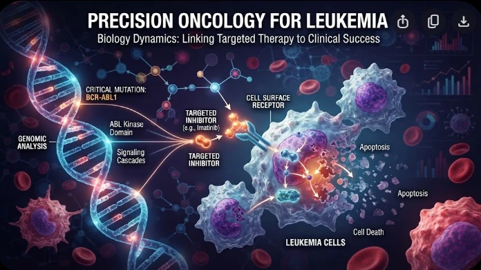
The article examines leukemia as an evolving population under targeted therapy, using chronic myeloid leukemia as the main example. BCR–ABL1 inhibition can produce deep responses, but every treatment changes the competitive environment among malignant clones. Quantitative transcript monitoring provides a continuous measure of disease burden, while deep sequencing can identify emerging resistance mechanisms before hematologic progression. A detected mutation is clinically meaningful only when its
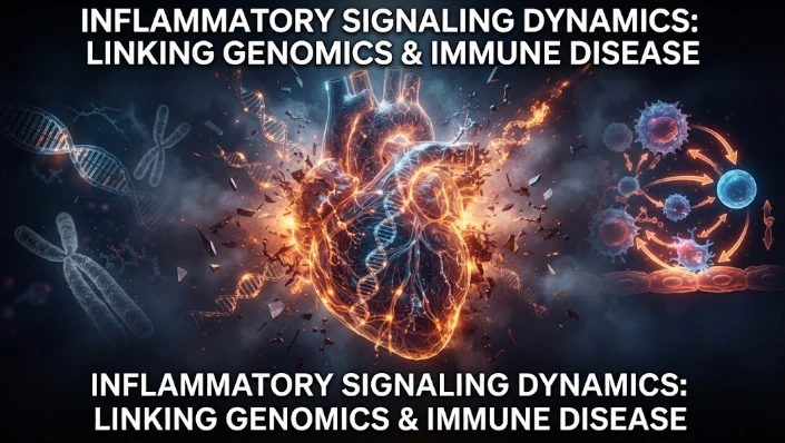
The article examines how cardiovascular genetic risk becomes biologically active through cell-specific inflammatory signaling. Because many vascular-risk variants lie in regulatory DNA rather than coding regions, their effects may appear only in particular endothelial, macrophage, smooth-muscle, or lymphocyte states. Single-cell studies of human atherosclerotic lesions and multiomic endothelial experiments are used to illustrate this context dependence. The article also discusses IL6-related sig
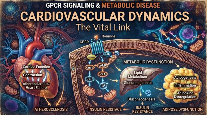
The article examines cardiovascular GPCR signaling as a dynamic response system whose abnormalities can remain hidden in resting measurements. Mechanical stretch, hormones, nutrients, sympathetic activity, and fluid balance can change the amplitude, duration, localization, and recovery of receptor signaling. Examples include mechanically activated angiotensin-receptor signaling, endosomal signaling, beta-arrestin-dependent trafficking, and biased glucagon-like peptide-one receptor responses. The