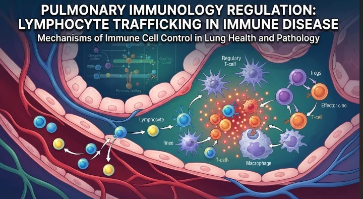
The article describes lymphocyte trafficking as a central regulator of pulmonary immunity because effective defense depends on where immune cells move, when they arrive, and how long they remain. It explains how chemokines, receptors, endothelial adhesion, integrins, tissue signals, and receptor desensitization guide lymphocytes from blood into airways and lung tissue. CXCR3-related pathways, IL-16-mediated receptor regulation, and histamine H4 signaling illustrate how migration can be selectively amplified or restrained. The article emphasizes that excessive trafficking can worsen asthma or immune-mediated injury, while insufficient recruitment can impair antiviral or antifungal defense. Single-cell, spatial, and paired blood–airway profiling are proposed to separate entry, proliferation, retention, and exit. Functional migration assays and tissue models are needed for mechanism. The article concludes that pulmonary therapy should restore an appropriate pattern of immune-cell traffic rather than simply increase or suppress total lymphocyte numbers.
The lung must protect a huge surface with only some of the inhaled particles causing harmful inflammation. Lymphocyte trafficking plays a key role in that balance, as immune cells need to be at the right tissue compartment, at the right stage of disease, and in the right activation state. The pulmonary immune system is not only dependent on the presence of the different lymphocytes but on how chemokines, adhesion molecules, receptors and local tissue signals guide their migration within the blood, airways, interstitium and lymphatic tissue.
The exit of T cells from the circulation involves a series of coordinated endothelial contact, chemokine sensing, activation of integrins, firm adhesion and tissue entry. Within the lung, further signals direct the migration to other target areas of the epithelium, inflammatory sites, or regulatory areas. Chemokine receptors serve as navigation systems which undergo changes depending on receptor expression, desensitization, ligand concentration and previous activation. Trafficking is seen, therefore, as a process which is regulated instead of being merely a movement to inflammation.
The importance of CXCR3 in pulmonary immune responses is illustrated. It binds to molecules that are often produced in response to interferon signaling, such as CXCL9, CXCL10, and CXCL11. The activated T cells with CXCR3 can then migrate along those gradients to the damaged tissues. Research on CXCL11 (also known as I T A C) confirmed its potent activity towards activated T cells. These routes link local cytokine production to the recruitment of effector lymphocytes.
The responsiveness to chemokines can also be selectively suppressed. Targeting of IL16 revealed that it can desensitize CXCR3 on T cells in a CD4 dependent manner. Thus a cell with the appropriate chemokine receptor could then become temporarily desensitized to its ligand. This kind of cross regulation makes it difficult to reduce trafficking models to the level of receptor abundance. A functional receptor state is important, particularly in the context of inflammation where there is a multitude of competing signals.
There's an extra layer: Histamine signaling. Recruitment of regulatory T cells has been shown in models of airway inflammation with studies on the histamine H4 receptor. Regulatory T cells with expression of FoxP3 have the ability to regulate excessive immune activation and may also modulate allergic disease. A mediator of allergy can thus be involved in cell migration, and in chemotaxis and lung immune regulation.
Both inadequate and excessive lymphocytes movement is relevant to these mechanisms; this is why it is relevant to pulmonary disease. The weak recruitment can be a cause of delayed clearance during viral/fungal infection and allow pathogen spread. Excessive recruitment in asthma or immune mediated lung injury can lead to a greater production of cytokines, secretion of mucus, tissue damage, or airway hyperresponsiveness. In one environment, a trafficking mechanism that promotes protection may be observed in another environment, it may promote pathologies.
Once in the tissues, lymphocytes undergo further changes in function. The environment in which a T cell finds itself in the vascular compartment is different from that in which a T cell resides close to airway epithelium. Lung tissue can store resident memory populations for an extended period following infection, and react very quickly to subsequent exposures. Regulatory populations may cluster at inflammation sites. Spatial organization is then part of immune phenotype.
That organization can be captured by modern pulmonary immunology, with approaches applied at a single cell and spatial scale. ST or multiplex imaging determines where the states occur, single cell RNA sequencing determines the states and receptor programs of lymphocytes. Systemic activation from local tissue recruitment can be distinguished through paired sampling of blood and airway. Longitudinal sampling can provide information on whether a cell population increases due to a proliferation rate, an increase in entry, a decrease in exit, or a prolonged cell residence time in a tissue.
Despite the limit on expression data, functional migration assays still are needed, as a lack of movement is not synonymous with a lack of expression. Cells can be challenged with known chemokine gradients and the receptor dependency can be identified by receptor blockade. Model of lung tissue and organotypic cultures can add endothelial/epithelial barrier. Animal models can be used to determine if modification of the trafficking receptor impacts disease severity, pathogen burden, airway physiology, or airway injury. These layers take a step towards mechanism.
In clinical translation, select the right candidates. Blocking a trafficking pathway may result in less inflammatory damage as well as less protective immunity. For instance, a therapy aimed at recruiting through CXCR3 might affect antiviral or antitumor responses not directly related to the disease. Strategies should aim to identify which subset of lymphocytes is targeted, which tissue compartment is altered, and what is the role of immune surveillance. Widespread suppression may alleviate one inflammatory issue, only to exacerbate another.
Determination of the right immune state can be made with the help of biomarkers. A combination of circulating chemokines, expression of receptors, airway cytokines, imaging signals and tissue transcriptional profiles might infer the disease process of excessive recruitment of effectors, insufficient regulation, or persistent tissue retention. This classification would be more useful than one inflammatory marker. This may also account for the clinical differences in response to immune modifying therapy in patients with identical clinical diagnosis.
In acute lung disease, temporal design is of particular importance. Early recruitment is protective; the continued infiltration after pathogen control can be a cause of injury. A treatment given at an inappropriate stage could have an adverse effect. Longitudinal immune profiling should thus be aligned with disease stage, pathogen load, therapeutic exposure and recovery from infection. That sequence can't be resolved by static comparison between sick and healthy groups.
A helpful model of pulmonary lymphocyte trafficking involves considering it as a regulated flow through interconnected compartments. Perspective and direction, receptors, entry control by endothelial barriers, local mediators and changes in response, persistence and function in tissue niches. Disease occurs when this flow is out of time, out of direction, and/or poorly controlled. This model can fit all the processes related to infection, allergy, autoimmunity and chronic inflammatory lung disease under one mechanistic umbrella.
Trafficking studies should be given more attention to endothelial biology. Pulmonary vessels are not just a "doorway. Cytokines change endothelial adhesion molecule properties, endothelial permeability, and presentation of chemokines leading to regional differences in cell entry. Therefore, even if the programs of the receptors of lymphocytes do not change, vascular injury can re-direct immune traffic. A combination endothelial and lymphocyte profiling can determine if abnormal recruitment takes place on side of the vessel or the immune cell side.
Another step that hasn't been extensively studied is Lymphatic exit. Cells that invade the lung tissue may return via the lymphatics or become locally resident. A lack of egress may create a chronic inflammatory state when recruitment signals are decreased. Two mechanisms of entry that lead to similar numbers of cells in the tissues, continuous entry and impaired departure can be distinguished by measuring markers in addition to chemokine receptors, and require different types of interventions.
Trafficking clinical trials should incorporate infection surveillance and vaccine response as safety endpoints. Pathogenic inflammation of the lungs can have a negative effect on immune responses in other parts of the body. Respiratory infection rates, viral clearance, and antigen specific memory can uncover unwanted immune suppression. This safety biology is particularly relevant to the therapy of chronic lung disease for long duration.
Apart from that, airway and parenchymal disease should be separated if possible. Bronchial dominant inflammation signals may be different to those active in alveolar or interstitial injury. Therefore, sampling from the blood or sputum only may result in the failure to capture a compartment specific mechanism. Imaging guided tissue analysis, when clinically appropriate, can link local chemokine programs to regional pathology and prevent attempts to treat the lung as a single immune organ.
The compartment aware approach can also enhance the interpretation of the biomarkers by connecting the circulation with the compartment that generates them instead of the assumption that all pulmonary inflammatory markers are derived from the same compartment.
The study of immune cells in motion rather than isolated labels allows for a more precise pulmonary immunology. Identifying tissue sources, the nature of signals that attract lymphocytes, the regulation of receptor sensitivity, and events following their entry into tissue can provide clues to intervention points that are not apparent from routine blood counts. The therapeutic aim is not just the reduction or augmentation of the number of lymphocytes. It's the reestablishment of the proper cellular traffic pattern in a given stage and biological context of lung disease.
CITE THIS ARTICLE
Tasmeem, S. (2026). Pulmonary Immunology Regulation through Lymphocyte Trafficking in Immune Disease. https://www.sadiaresearchhub.com/article/76d3ff68-e9a1-538a-8e7c-147e6131175c
S. Tasmeem, “Pulmonary Immunology Regulation through Lymphocyte Trafficking in Immune Disease,” 2026. [Online]. Available: https://www.sadiaresearchhub.com/article/76d3ff68-e9a1-538a-8e7c-147e6131175c
Tasmeem, S. (2026) ‘Pulmonary Immunology Regulation through Lymphocyte Trafficking in Immune Disease’. Available at: https://www.sadiaresearchhub.com/article/76d3ff68-e9a1-538a-8e7c-147e6131175c
Tasmeem, Sadia. “Pulmonary Immunology Regulation through Lymphocyte Trafficking in Immune Disease.” 2026, https://www.sadiaresearchhub.com/article/76d3ff68-e9a1-538a-8e7c-147e6131175c.
Sadia Tasmeem. “Pulmonary Immunology Regulation through Lymphocyte Trafficking in Immune Disease.” (2026). https://www.sadiaresearchhub.com/article/76d3ff68-e9a1-538a-8e7c-147e6131175c.
Tasmeem S. Pulmonary Immunology Regulation through Lymphocyte Trafficking in Immune Disease. 2026. https://www.sadiaresearchhub.com/article/76d3ff68-e9a1-538a-8e7c-147e6131175c
@misc{article_76d3ff68e9a1538a8e7c147e6131175c,
  title = {Pulmonary Immunology Regulation through Lymphocyte Trafficking in Immune Disease},
  author = {Sadia Tasmeem},
  year = {2026},
  url = {https://www.sadiaresearchhub.com/article/76d3ff68-e9a1-538a-8e7c-147e6131175c},
}
TY  - GEN
AU  - Sadia Tasmeem
TI  - Pulmonary Immunology Regulation through Lymphocyte Trafficking in Immune Disease
PY  - 2026-06-14
UR  - https://www.sadiaresearchhub.com/article/76d3ff68-e9a1-538a-8e7c-147e6131175c
ER  -
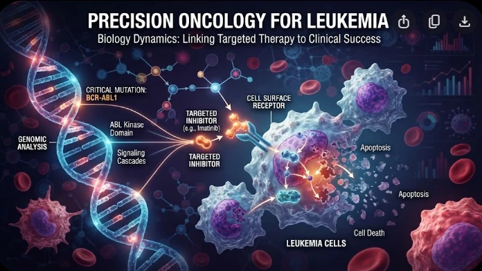
The article examines leukemia as an evolving population under targeted therapy, using chronic myeloid leukemia as the main example. BCR–ABL1 inhibition can produce deep responses, but every treatment changes the competitive environment among malignant clones. Quantitative transcript monitoring provides a continuous measure of disease burden, while deep sequencing can identify emerging resistance mechanisms before hematologic progression. A detected mutation is clinically meaningful only when its
The article examines how cardiovascular genetic risk becomes biologically active through cell-specific inflammatory signaling. Because many vascular-risk variants lie in regulatory DNA rather than coding regions, their effects may appear only in particular endothelial, macrophage, smooth-muscle, or lymphocyte states. Single-cell studies of human atherosclerotic lesions and multiomic endothelial experiments are used to illustrate this context dependence. The article also discusses IL6-related sig
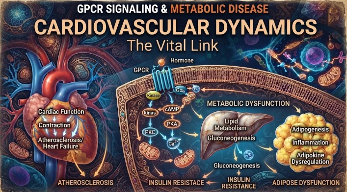
The article examines cardiovascular GPCR signaling as a dynamic response system whose abnormalities can remain hidden in resting measurements. Mechanical stretch, hormones, nutrients, sympathetic activity, and fluid balance can change the amplitude, duration, localization, and recovery of receptor signaling. Examples include mechanically activated angiotensin-receptor signaling, endosomal signaling, beta-arrestin-dependent trafficking, and biased glucagon-like peptide-one receptor responses. The