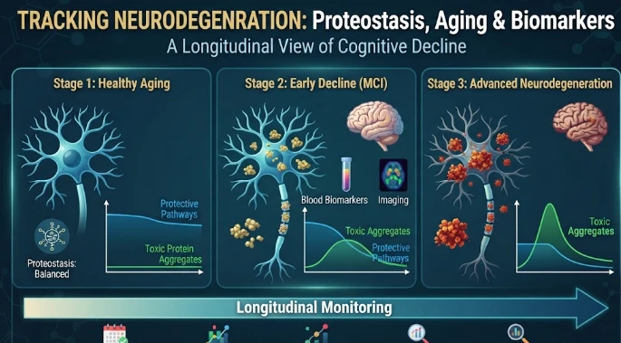
The article describes neurodegeneration as a progression from age-related proteostatic stress to cell-specific and eventually circuit-level failure. Neurons face long-term burdens in protein folding, trafficking, degradation, mitochondrial function, and organelle quality, while disease-associated proteins such as tau, alpha-synuclein, TDP-43, and polyglutamine expansions exploit different vulnerabilities. Longitudinal biomarkers can help order pathology-specific changes, glial responses, axonal injury, imaging abnormalities, and functional decline over time. The article stresses that selective neuronal vulnerability, vascular and metabolic aging, sleep, immune changes, and mitochondrial stress all modify progression. Patient-derived neural models and perturbation screens can test whether chaperone, autophagy, lysosomal, or stress pathways are causal and reversible. Spatial proteomics can identify local neuron–glia interactions and possible propagation mechanisms. Clinically useful markers should detect modifiable transition points before irreversible neuronal loss. The central goal is to identify windows where restoring proteostatic reserve can still alter the course of degeneration.
Most often the signs of neurodegeneration are observed once the function has already diminished, but the molecular processes that cause it can be underway for many years. Proteostasis offers a good bridge from age-related early biology to later tissue loss, as neurons rely on the continuous control of folding, trafficking, degradation and organelle quality of proteins. Progression occurs when the number of damaged proteins and cellular stress is greater than the ability of these compensatory systems and varies from cell to cell and person to person.
As people get older, the chances of reaching that threshold increase. Protein turnover shifts, autophagic function may be reduced, lysosomal function is less reliable, mitochondrial damage accumulates and stress signaling may persist longer. These properties are not part and parcel of neurodegeneration. The risk lies in interaction: aggregated burden due to impaired clearance, stress of aggregates on mitochondria and transport systems, and energy failure further compromises quality control.
Different types of disease associated proteins take advantage of those weaknesses. Tau can alter the biology of the axon and synapse, α synuclein can influence on vesicle and membrane biology, TDP 43 pathology can influence RNA handling, and expanded polyglutamine proteins can overwhelm the folding machineries. Each are molecularly distinct, but both cause a long-term problem that cannot be solved by the rapid division of cells to dilute damage away.
Progressions show up in areas of selective vulnerability. The metabolic demands, axonal architecture, firing patterns and calcium loads of dopaminergic neurons, motor neurons, cortical projection neurons and others are different. What is ok in one cell type may be a disaster in another. Single cell analysis is thus required to explain the regionally specific degeneration of a disease protein, the gene encoding which is widely expressed.
Longitudinal biomarkers can serve as markers for various levels of this process. These phosphorylated tau species may be associated with specific protein pathology in disease, while the neurofilament light may represent axonal injury, and the glial fibrillary acidic protein may represent astrocytic response. Repeated measurements determine if the protein pathology marker is increasing before the injury marker or if there is an early detectable change along with a glial activation. Temporal ordering assists to differentiate upstream events and downstream damage.
Those trajectories can be altered by the aging process of biology. Vascular dysfunction can impair nutrient and oxygen delivery, metabolic disease can lead to oxidative and inflammatory stress and sleep disruption to protein clearance and neuronal recovery. With immune aging, the behavior of microglia and the tone of cytokines change. The crosstalk between organs should be a part of the study of a proteostasis pathway, not a stand-alone neuronal pathway.
Patient generated neural models can test causal links suggested by longitudinal cohorts. A disease associated variant can be introduced into an iPSC and the iPSC differentiated to a neuron or glia and compared with a gene corrected isogenic iPSC. To test the effect of correcting one component, a cell's function could be revealed by perturbing the autophagy, chaperones, pathways to the lysosomes or stress regulators. Rescue is more convincing than seeing a pathway as being "abnormal.
One of the difficulties faced by such models is maturation. Cells grown in culture tend to be more juvenile than cells during the aging process; most neurodegenerative diseases happen after a number of decades of aging. Researchers have the ability to add oxidative stress, metabolic stress, proteotoxic stress, or inflammatory stress but the induced stress is not the same as natural aging. Mechanisms that are also found in human tissue are the most credible findings, as are longitudinal biomarker data.
Perturbation screening will detect points at which progression is still reversible. Genetic screens can deplete the expression of candidate regulators, small molecule screens can manipulate proteostasis pathways. Rescue of protein aggregation must also enhance mitochondrial function or synaptic physiology or cell survival to be mechanistically informative. Some visible markers can be suppressed without restoration of function, masking disease biology instead of correcting it.
Spatial proteomics can reveal the propagation within a tissue compartment. Protein pathology frequently interacts with glial responses, vascular cells, and ECM. Local feedback circuits can be determined by mapping damaged neurons in close proximity to activated microglia and/or astrocytes. Changes in proteins, chaperones and inflammatory mediators in regions can help identify the mechanisms that lead to differences in circuit deterioration.
Future clinical biomarker testing needs to be associated with clear outcomes. A candidate marker can be repeated many times prior to cognition, movement and imaging or electrophysiology changes. The predictive value should be evaluated in independent sets and in various disease stages. A marker that is useful for early detection is not necessarily useful for monitoring treatment; a marker that tracks injury is not necessarily a marker of the causal proteostasis mechanism.
When writing progression models, competing explanations should also be included. There are various neurological conditions in which the NF-L level can increase, and inflammation proteins may fluctuate in systemic diseases. Circulating concentrations may be affected by kidney function and age. Disease-specific markers with general injury / aging markers can be useful for interpretation and if serial measures are used, and they show consistent changes across different markers.
In the field of clinical translation, a decision point is needed. The most useful time for biomarkers is when they detect a point at which an intervention might be able to make a difference. The earlier irreversible neuronal loss can be detected before a modifiable mechanism, the better the prognosis, than therapy. Proteostasis research is focused on pushing the signal up the line towards the reducing cellular reserve before it's entrenched and widespread degeneration is the norm.
There can also be a propagation of proteostasis failure between cells. Misfolded protein aggregates can be transported between neurons, and inflammatory signals from glia can alter protein clearance in neighbouring cells. The evidence of templated spread is clear in a few proteinopathies and experimental systems, with tissue progression as both network transmissions and cell autonomous stresses being modeled.
Microglia can have a protective or damaging effects based on stage. Phagocytic responses may be beneficial in the early stages to clear debris accumulated in the lesion, but chronic activation may produce inflammatory mediators that would exacerbate neuronal stress. This may mean that the same longitudinal glial biomarkers may have different interpretations during progression. A high signal may indicate compensation and/or permanent injury.
Such stage differences can be identified with combination biomarkers. A pathway formulated by a combination of a pathology specific marker, an axonal injury marker, and a glial response marker is a multidimensional trajectory. Increase in pathology followed by glial activation and increase in neurofilaments later, is compatible with a model other than simultaneous changes. Multiple imaging can offer an anatomical snapshot of the molecular timeline.
Intervention experiments should involve testing for durability after the perturbation has been removed. A compound that only decreases aggregates while present is different than a compound that can restore a self sustaining quality control state. Neural models can be washed out and the stability of proteostasis can be measured. If indeed a regulatory network, rather than just a single protein concentration, had been reset, then durable rescue would come into consideration.
Loss of network compensation can also be used to model progression. Early disease can lead to alternative pathways that maintain the output of synapses despite molecular stress. Functional decline starts when there is a lack of compensation. Longitudinal physiology such as electrophysiology or cognitive testing can be correlated with molecular markers to pinpoint that tipping point.
A progression signature should also help identify how much disability is the result of active degeneration and how much is the result of accumulated disability. After a lot of neuronal loss, biomarkers can drop. Therefore, imaging and functional context is necessary to interpret late stage decline, not the assumption of a decrease in concentration equates to biological recovery.
These tipping points can be useful parameters for a prevention study which aims to maintain function before the circuits are lost.
This progression framework is supported by Richard I. Morimoto's research on proteostasis, aging and neurodegenerative disease. Longitudinal biomarkers can help identify the time when molecular stress is starting to accelerate, and patient derived models can help test which changes are causal and reversible. The strongest translational value is achieved when three scales of aging related loss of protein quality control, cell type specific vulnerability, and measurable progression in living humans are linked together. Neurodegeneration subsequently is a cascade of maladaptations that make appreciable windows apparent for where recovering proteostatic capacity will be most important.
This time can inform trials that focus on reserve maintenance.
CITE THIS ARTICLE
Tasneem, S. (2025). Neurodegeneration Progression through Proteostasis and Aging Biology Using Longitudinal Biomarkers. https://www.sadiaresearchhub.com/article/92be64d6-efc2-54e1-a9d5-4dba41dc6cf5
S. Tasneem, “Neurodegeneration Progression through Proteostasis and Aging Biology Using Longitudinal Biomarkers,” 2025. [Online]. Available: https://www.sadiaresearchhub.com/article/92be64d6-efc2-54e1-a9d5-4dba41dc6cf5
Tasneem, S. (2025) ‘Neurodegeneration Progression through Proteostasis and Aging Biology Using Longitudinal Biomarkers’. Available at: https://www.sadiaresearchhub.com/article/92be64d6-efc2-54e1-a9d5-4dba41dc6cf5
Tasneem, Sadia. “Neurodegeneration Progression through Proteostasis and Aging Biology Using Longitudinal Biomarkers.” 2025, https://www.sadiaresearchhub.com/article/92be64d6-efc2-54e1-a9d5-4dba41dc6cf5.
Sadia Tasneem. “Neurodegeneration Progression through Proteostasis and Aging Biology Using Longitudinal Biomarkers.” (2025). https://www.sadiaresearchhub.com/article/92be64d6-efc2-54e1-a9d5-4dba41dc6cf5.
Tasneem S. Neurodegeneration Progression through Proteostasis and Aging Biology Using Longitudinal Biomarkers. 2025. https://www.sadiaresearchhub.com/article/92be64d6-efc2-54e1-a9d5-4dba41dc6cf5
@misc{article_92be64d6efc254e1a9d54dba41dc6cf5,
  title = {Neurodegeneration Progression through Proteostasis and Aging Biology Using Longitudinal Biomarkers},
  author = {Sadia Tasneem},
  year = {2025},
  url = {https://www.sadiaresearchhub.com/article/92be64d6-efc2-54e1-a9d5-4dba41dc6cf5},
}
TY  - GEN
AU  - Sadia Tasneem
TI  - Neurodegeneration Progression through Proteostasis and Aging Biology Using Longitudinal Biomarkers
PY  - 2025-07-20
UR  - https://www.sadiaresearchhub.com/article/92be64d6-efc2-54e1-a9d5-4dba41dc6cf5
ER  -
The article examines leukemia as an evolving population under targeted therapy, using chronic myeloid leukemia as the main example. BCR–ABL1 inhibition can produce deep responses, but every treatment changes the competitive environment among malignant clones. Quantitative transcript monitoring provides a continuous measure of disease burden, while deep sequencing can identify emerging resistance mechanisms before hematologic progression. A detected mutation is clinically meaningful only when its
The article examines how cardiovascular genetic risk becomes biologically active through cell-specific inflammatory signaling. Because many vascular-risk variants lie in regulatory DNA rather than coding regions, their effects may appear only in particular endothelial, macrophage, smooth-muscle, or lymphocyte states. Single-cell studies of human atherosclerotic lesions and multiomic endothelial experiments are used to illustrate this context dependence. The article also discusses IL6-related sig
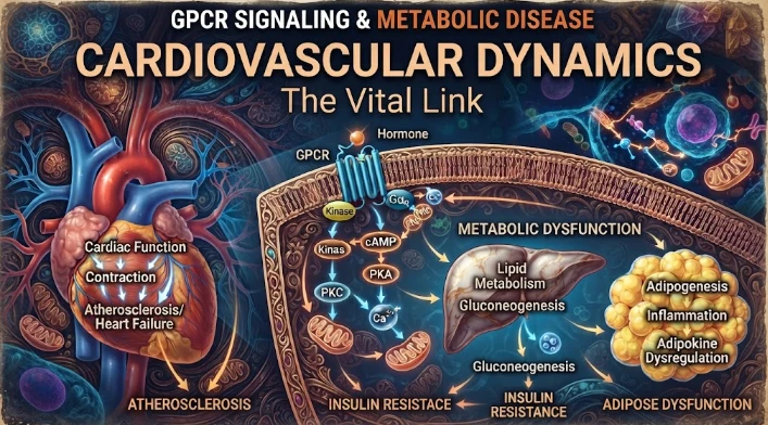
The article examines cardiovascular GPCR signaling as a dynamic response system whose abnormalities can remain hidden in resting measurements. Mechanical stretch, hormones, nutrients, sympathetic activity, and fluid balance can change the amplitude, duration, localization, and recovery of receptor signaling. Examples include mechanically activated angiotensin-receptor signaling, endosomal signaling, beta-arrestin-dependent trafficking, and biased glucagon-like peptide-one receptor responses. The