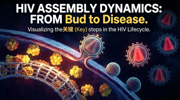
The article focuses on the kinetics of HIV assembly, describing particle formation as a sequence of molecular transitions rather than a single budding event. Gag accumulates at discrete membrane sites, viral RNA is captured, the immature lattice grows, ESCRT proteins are recruited transiently, membrane fission occurs, and protease-driven maturation reorganizes the particle. Live-cell studies show that these steps unfold over minutes and that delayed budding can reduce infectivity by allowing maturation to occur in the wrong structural context. The article emphasizes single-particle analysis because average release measurements can hide stalled assembly events. Perturbing Gag interactions with TSG101, ALIX, can place candidate proteins within the kinetic sequence. Proximity labeling and proteomic approaches can identify additional assembly-site components for temporal testing. The central message is that timing itself is a determinant of viral fitness, and interventions should be evaluated for effects on assembly, release, maturation, and final infectivity.
Once Gag gets to the membrane, HIV will not leave. A series of molecular transitions has been observed in living cells that are involved in viral assembly. Gag is concentrated at specific membrane locations, viral RNA is grabbed, the immature lattice expands, ESCRT proteins arrive in a transient fashion, membrane fission takes place and the protease-driven maturation reorganizes the particle. There is a specific timing to each transition and the timing of the infectivity is related to coordinating transitions.
Many live cell assembly measurements start with the accumulation of gags. The fluorescently labeled Gag appears in distinct clusters of the plasma membrane, which increase in size over the course of minutes. Single clusters typically release 1 virion and not a continuous release from a single permanent factory. This is consistent with the nucleation and growth model, where new particle growth occurs at what are thought to be randomly selected membrane locations.
Those sites are partially determined by the composition of the membrane. Gag is specific for phosphoinositide lipids and can bind and localize phosphatidylinositol bisphosphate and cholesterol. The assembling virus, then, modifies the physical environment of the membrane as it assembles the lattice. Lipid organization can modulate curvature, protein mobility and/or recruitment of other viral components.
Viral RNA is early player. This packaging signal is recognized by Gag and RNA is able to nucleate and/or stabilize assembly. Self assembly of Gag has been observed to occur selectively when experiments were conducted with reconstituted membrane models. The mutations may be subtle and cause a decrease in RNA specificity, so genome choice is coupled with structural growth.
In addition, a cellular RNA is introduced into the process. Host 7SL RNA is recruited at or near the time of Gag association to the plasma membrane, and RNA binding proteins like the Staufen1 bind to viral RNP complexes. The assembly site on the other hand may be a protein-rich environment of RNA rather than a purified viral machine.
ESCRT recruitment is delayed and is transient. TSG101 can bind Gag via its PTAP late domain, and ALIX offers another pathway toward ESCRT III machinery. The live imaging revealed that later components come after the Gag accumulation, fitting the idea that later components are involved in the constriction and cutting of the narrow membrane neck connecting the budding virion to the cell.
This timing is important for the function. Delaying release can be achieved by experiments that disrupt the Gag interaction with TSG101 or ALIX.Experiments disrupting the Gag interaction with TSG101 or ALIX can delay release. If budding is delayed too long, viral protease can be activated before the particle is sufficiently separated, allowing missing parts of the viral particle to escape or viral components to mature in the wrong structural environment. Even after a particle finally exits the membrane its infectivity is reduced.
This results in another fast structural change during maturation. The Gag and Gag Pol proteins are precursors that are cleaved to form the mature capsid proteins by the protease. Fight the protease inhibitors are an example of drugs that inhibit this rearrangement. An immature particle that has been released is not a fully infectious virion.
Change in host restriction affects the release dynamics of also. Tetherin plays a role in retaining budded particles at the surface of cells, thus effectively preventing successful membrane scission from becoming a failed dissemination event. Vpu counters tetherin, indicating that viruses must also overcome active host defenses to be released, in addition to the machinery necessary to assemble them. The strength of this restriction can be modified by interferons.
Super resolution microscopy provides additional spatial information, in addition to the temporal one. ESCRT proteins are found in the budding neck, but not across the whole virion surface. This geometry is applicable to a membrane fission mechanism that takes place at a confined region at the viral-cell interface. So structural imaging can then be used to test the validity of the kinetic observations in terms of their physical location.
Total viral protein should not be the only measure of quantitative analysis. Different defects may be detected in the number of assembly sites, the growth rate of Gag clusters, the delay to ESCRT recruitment, the time before release and infectivity of released particles. A perturbation that diminishes the total release may occur at the nucleation step, the membrane binding step, the scission step, the maturation step or even at a preceding step of viral expression.
These stages can be mapped with the aid of perturbation experiments. Late domains are mutated to test ESCRT dependence, membrane targeting is altered to test Gag localization, RNA packaging signals are changed to test genome recruitment, and host factors are mutated to test cellular support. If a delay is observed upon rescue with wild type components, it would be more convincing that the delay is due to the intended pathway.
Single particle analysis can expose variation that is not apparent in the averages. Some assembly sites develop normally and others stall or recruit an abnormal amount of ESCRT. Subpopulations associated with subpopulation-specific distributions of assembly time and release delay can be identified and used to measure infectivity loss. Even if a small proportion of particles are very defective the mean value can hardly change.
Fluorescent reporters, whose signal is altered following the action of the protease, can be used for studies of protease timing. A perturbation could be aligned to either the accumulation or release of Gag, allowing the determination of whether a perturbation promotes maturation or slows down the fission. This evidence of time is direct and is more robust than evidences of time based on endpoint particle morphology.
Membrane diffusion and trafficking are energy-dependent processes and can be affected by temperature and cell metabolism, which can affect assembly kinetics. These conditions should be noted on controlled experiments. Apparent differences among cell lines may be due to state of growth or cell membrane composition rather than to a specific host factor.
Another kinetic checkpoint is the dimerisation of the viral RNA. HIV encapsulates two copies of the genome, and structural modifications in the vicinity of the packaging region affect dimerization and Gag recognition. Mutations can thus influence the assembly even in the presence of normal total RNA levels.
A full kinetic model should include a link between rate changes and fitness. The length of time required for assembly does not necessarily correlate with any negative consequences if particles are still infectious, and a brief delay at an important late stage can significantly decrease infectivity. By aligning live cell timing with downstream infection assays, the transitions that are demonstrated to be the rate limiting steps can be identified.
Structural capsid characteristics are used to quantitatively measure particle maturation. Cryogenic tomography can differentiate between immature lattices and mature conical cores, and may be capable of identifying defects in a population. When used in combination with infectivity, structural classification can be used to determine which abnormal morphology is likely to affect function.
The proteomic profiling of the assembly site composition is also possible. Proximity labeling can be used to identify Gag interacting proteins in specific time periods. Candidates then can be individually perturbed and inserted into the kinetic sequence with live imaging. This is a strategy that combines discovery and temporal mechanism.
Alternative pathways can be revealed with cell type comparison. Differences in membrane organisation and trafficking between macrophages and T cells mean that a factor used in one context is partially redundant in another. The antiviral breadth of the activity should be confirmed in the primary cellular reservoirs and targets of infection.
Heterogeneity of the kinetic can be a drug target. A compound that selectively extends the scission step may lead to a significant fraction of particles being beyond the safe margin for maturation even if it does not inhibit budding. This method would not be captured by end-point assays that only measured the total number of particles. Time resolved imaging can thus show antiviral activity which static measurements underestimate.
The most informative experiments thus link single particle timing, maturation of the structure, genome content, and downstream infectivity into the same mechanistic framework.
In the field of HIV assembly and host virus interactions, Carol A. Carter's research highlights the need to study viral budding as an ongoing process and not as an endpoint. All of this requires the virus to orchestrate membrane selection, RNA capture, the formation of the lattice, the recruitment of host machines, the fission and the maturation in a tight temporal window. A study of infectious disease may be able to determine vulnerable transitions by quantifying the impact of certain perturbations on the sequence. Not all viral protein must be eliminated, and even targeting one critical step in one of the assembly transitions could be sufficient to stop a particle from being infectious.
This integrated design is capable of detecting vulnerable time windows that are not detected in bulk release assays and can discriminate between delayed assembly and immature maturation.
The phenomenon of timing is still explanatory.
CITE THIS ARTICLE
Tasneem, S. (2025). Viral Assembly Dynamics Linking HIV Assembly to Infectious Disease. https://www.sadiaresearchhub.com/article/a21348ff-3a2c-5946-85e5-dc61ab83825e
S. Tasneem, “Viral Assembly Dynamics Linking HIV Assembly to Infectious Disease,” 2025. [Online]. Available: https://www.sadiaresearchhub.com/article/a21348ff-3a2c-5946-85e5-dc61ab83825e
Tasneem, S. (2025) ‘Viral Assembly Dynamics Linking HIV Assembly to Infectious Disease’. Available at: https://www.sadiaresearchhub.com/article/a21348ff-3a2c-5946-85e5-dc61ab83825e
Tasneem, Sadia. “Viral Assembly Dynamics Linking HIV Assembly to Infectious Disease.” 2025, https://www.sadiaresearchhub.com/article/a21348ff-3a2c-5946-85e5-dc61ab83825e.
Sadia Tasneem. “Viral Assembly Dynamics Linking HIV Assembly to Infectious Disease.” (2025). https://www.sadiaresearchhub.com/article/a21348ff-3a2c-5946-85e5-dc61ab83825e.
Tasneem S. Viral Assembly Dynamics Linking HIV Assembly to Infectious Disease. 2025. https://www.sadiaresearchhub.com/article/a21348ff-3a2c-5946-85e5-dc61ab83825e
@misc{article_a21348ff3a2c594685e5dc61ab83825e,
  title = {Viral Assembly Dynamics Linking HIV Assembly to Infectious Disease},
  author = {Sadia Tasneem},
  year = {2025},
  url = {https://www.sadiaresearchhub.com/article/a21348ff-3a2c-5946-85e5-dc61ab83825e},
}
TY  - GEN
AU  - Sadia Tasneem
TI  - Viral Assembly Dynamics Linking HIV Assembly to Infectious Disease
PY  - 2025-05-04
UR  - https://www.sadiaresearchhub.com/article/a21348ff-3a2c-5946-85e5-dc61ab83825e
ER  -
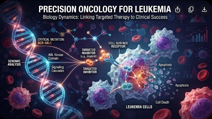
The article examines leukemia as an evolving population under targeted therapy, using chronic myeloid leukemia as the main example. BCR–ABL1 inhibition can produce deep responses, but every treatment changes the competitive environment among malignant clones. Quantitative transcript monitoring provides a continuous measure of disease burden, while deep sequencing can identify emerging resistance mechanisms before hematologic progression. A detected mutation is clinically meaningful only when its
The article examines how cardiovascular genetic risk becomes biologically active through cell-specific inflammatory signaling. Because many vascular-risk variants lie in regulatory DNA rather than coding regions, their effects may appear only in particular endothelial, macrophage, smooth-muscle, or lymphocyte states. Single-cell studies of human atherosclerotic lesions and multiomic endothelial experiments are used to illustrate this context dependence. The article also discusses IL6-related sig
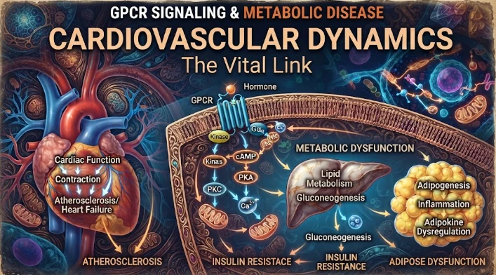
The article examines cardiovascular GPCR signaling as a dynamic response system whose abnormalities can remain hidden in resting measurements. Mechanical stretch, hormones, nutrients, sympathetic activity, and fluid balance can change the amplitude, duration, localization, and recovery of receptor signaling. Examples include mechanically activated angiotensin-receptor signaling, endosomal signaling, beta-arrestin-dependent trafficking, and biased glucagon-like peptide-one receptor responses. The