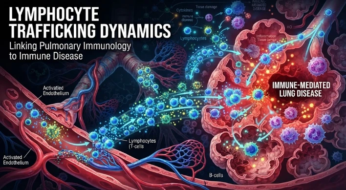
The article examines lymphocyte trafficking as a dynamic process that shapes pulmonary inflammation and immune disease. Activated lymphocytes interact with pulmonary endothelium, follow chemokine gradients, activate integrins, enter tissue, and then undergo further regulation through retention or egress. CXCR3 and its interferon-inducible ligands are discussed alongside IL-16-mediated receptor desensitization and histamine H4 signaling, showing that receptor expression does not necessarily equal migratory competence. Once inside the lung, lymphocytes can become resident, persist locally, or support protective and pathogenic responses depending on disease context. The article recommends longitudinal paired sampling of blood, airway, and tissue, combined with single-cell states, clonotypes, spatial profiling, migration assays, and cell-tracking methods. It also stresses distinguishing recruitment from local proliferation or failed exit. Therapeutic targeting must preserve antimicrobial surveillance. Overall, the article frames lung immune disease as a problem of abnormal cellular flow, timing, direction, and persistence.
Lymphocytes are not merely wandering in to the diseased lung. The sequence of chemokine gradients, endothelial signals, adhesion events, receptor desensitization, tissue retention, and exit pathways are constantly changing during the process of movement. Protection and pathology can be magnified at each stage. This is thus a problem of cellular geography: which populations of lymphocytes are found in each compartment and for how long are they activated?
The process starts in the circulation where activated lymphocytes interact with the inflamed pulmonary endothelium. Initial contact is promoted by selectins and integrins are activated by chemokines, allowing for firm adhesion and transendothelial migration. The expression of chemokine receptors restricts the cells that can respond to a specific chemokine. This selectivity enables the recruitment of different subsets of lymphocytes such as effector T cells, regulatory T cells, or other subsets, depending on the cytokine milieu at the inflamed site.
CXCR3 is emerging as an important marker of activated T cell trafficking. Interferon induced ligands (e.g. CXCL9, CXCL10, CXCL11) can generate gradients in inflammatory tissue. CXCL11 is particularly active for activated lymphocytes and contributed to the definition of the role of the inflammatory chemokines in guiding immune cells to sites of immune activation. Increased production of the ligands may amplify the local recruitment, but the function of the receptor is still modifiable after the cell receives some more signals.
Interleukin sixteen exhibits such adaptation. Desensitization of CXCR3 by CD4 dependent signaling could lead to decreased responsiveness. That indicates the limitations of using surface expression as a measure of trafficking competence. It is possible for a receptor to be on a lymphocyte with no corresponding gradient following. A large number of the mediators that mediate pulmonary inflammation are common to all of them, and thus the history of the receptor can be as important as its abundance.
Histamine related pathways can also direct immune traffic. Experimental studies have shown that activation of the H4 receptor is associated with recruitment of FoxP3 positive regulatory T cells in airway inflammation. This recruitment can help to reduce hyperactive immune responses, directly involving chemotaxis in the process of immune tolerance. So a mediator with allergic properties may modulate the effector inflammation/regulatory balance by selectively moving cells.
After entering the lung, the trafficking dynamics change. A small proportion of lymphocytes persist in the direction of airway epithelium, remaining close to blood vessels or lymphoid aggregates, or acquiring "resident memory" properties. Rapid responses during later exposure, but can sustain chronic inflammation, due to tissue residency. Thus, signals that regulate retention, survival and egress can be as critical as those that regulate entry.
Recruitment is beneficial to the disease context. Rapid arrival of antigen specific T cells can assist in clearance during the acute viral infection. In allergic asthma, recruitment of activated populations of helper T cells can exacerbate airway inflammation. In autoimmune or postinfectious insults, the initial cause may have resolved, but the cytokine-producing lymphocytes may persist. There is the possibility that different consequences can follow the same chemokine pathways in these states.
Static measurements are difficult to interpret because of the timing issue. High levels of CXCL10 in one phase may reflect productive antiviral recruitment; high levels of CXCL10 in recovery may represent ongoing inflammation that has not been resolved. The blood sample will not be able to determine if cells have invaded tissue, stuck in the pulmonary vasculature or if they have taken root in the tissue. Movement is best captured by longitudinal paired sampling of blood, airway material and tissue.
Single cell methods are able to determine the transcriptional state of migrating populations. Distinct trafficking programs can be defined by the expression of chemokine receptors, integrins, activation markers, exhaustion markers, and tissue residency genes. Spatial profiling adds the missing coordinate, revealing the spatial distribution of those states. It may be possible to differentiate between a population enriched near damaged epithelium and a similar population around vessels or lymphoid structures by using both methods together.
There are approaches, known as cell tracking, that can provide direct evidence. Estimates of entry, movement, and persistence can be obtained through experimental labeling, intravital imaging, photoconversion systems, and adoptive transfer models. Temporal changes, paired compartments, receptor phenotyping, and clonal sequencing are used to a greater extent in human studies. When interpreted in conjunction with the activation state and sampling time, T cell receptor clonotypes that are detected in blood and subsequently in airway samples can provide support for inference of recruitment.
Competing explanations should be tried using mechanistic experiments. Increased lung T cells may be indicative of increased recruitment, increased local proliferation, decreased apoptosis, or increased failure of migration out of the lung. In a model, blocking a CCR can provide an indication of the importance of CCR entry. After-arrival proliferation can be detected with proliferation markers and lineage tracing. These tests are essential to not mistake an increase in cell numbers for increased trafficking.
Therapeutic targeting should maintain protective immunity. General inhibition of lymphocyte traffic increases the vulnerability to anti-viral, anti-fungal, or anti-tumour mechanisms. A more precise intervention might occur if a receptor is present in high amounts on pathogenic cells, if the receptor is present on pathogenic cells only during certain stages of the disease, or if local chemokine production is altered. Any treatment based on biomarkers would have to be able to detect the trafficking state and confirm the maintenance of beneficial immune surveillance.
The molecular signals can be linked to the clinical outcomes using a systems model of pulmonary trafficking. There are three binding properties of chemokines: directionality, sensitivity (via changes in the state of chemokine receptors), and persistence (via changes in the activation status of the endothelium). These factors can be combined with lung function, imaging, pathogen burden, and symptoms to identify the movement pattern that is associated with recovery and which is predictive of ongoing disease.
Metabolism can act on trafficking following binding to receptor. The changes in glucose usage, mitochondrial processes, and lipid metabolism that take place in activated lymphocytes affect motility and persistence. Oxygen deprived regions of the lung may create a preference for other cell programs than oxygenated ones. Combining metabolic measurement with chemokine receptor state might help account for the difference in movement and/or survival of two phenotypically alike T cells within inflamed tissue.
Clonal information should be used to differentiate repeated recruitment from local persistence. Clones of T cells can be monitored in both the blood and lung compartments for months. If a particular clone is present in multiple airway samples, but not in blood, it could be either due to tissue retention or local growth. Blood and tissue clones that rise first are better evidence for active recruitment. The combination of clonotypes with single cell state, further improves interpretation.
Trafficking models also need to incorporate competition among chemokines. A single ligand is not likely to be expressed in inflamed lungs and a single receptor is not likely to be expressed on a single lymphocyte. Signals can amplify, counteract or neutralize each other. Competing gradient-based microfluidic migration assays can assess cell choices for gradients, provide a more physiological representation of tissue migration than assays focused on a single chemokine.
An intervention should have a trafficking phenotype that can be measured for clinical translation. The most rational use of a receptor antagonist is when the beneficial population utilizes an alternate pathway, and pathogenic cells have a strong dependence on the receptor. The eligibility signature might be receptor function, spatial distribution and disease stage. Measuring the same cells again after treatment might provide evidence of cell movement in the desired compartment and not just a decrease in a circulating biomarker.
This model can be further refined by the use of tissue exit markers and recirculation patterns. Cells can return to circulation or stay in the lung; this is affected by sphingosine related pathways, access to lymphatics, and residency programs. The measurement of these signals with entry receptors can help avoid misclassifications of a persistent population as continuous recruitment. This is important because as the cells are already present within a pathogenic niche, blocking entry does not remove these cells.
This comprehensive picture of entry, retention and exit can transform a snapshot of inflammation into a mechanistic description of cellular movement throughout the entire pulmonary response.
The central concept of pulmonary immunology is made more straightforward by replacing the concept of abundance of lymphocytes by the concept of flow of lymphocytes. Four types of recruitment can lead to immune disease: too much, too late, wrong direction, and too long. Circulation to tissue and exit can be mapped for identification of points at which there is a failure to regulate. This dynamic view helps to develop a better base for therapies aimed at modifying inflammation without compromising the immune functions that are essential for pulmonary defense.
CITE THIS ARTICLE
Tasmeem, S. (2026). Lymphocyte Trafficking Dynamics Linking Pulmonary Immunology to Immune Disease. https://www.sadiaresearchhub.com/article/a7a67448-bcfc-5a0b-aeb8-966219cec460
S. Tasmeem, “Lymphocyte Trafficking Dynamics Linking Pulmonary Immunology to Immune Disease,” 2026. [Online]. Available: https://www.sadiaresearchhub.com/article/a7a67448-bcfc-5a0b-aeb8-966219cec460
Tasmeem, S. (2026) ‘Lymphocyte Trafficking Dynamics Linking Pulmonary Immunology to Immune Disease’. Available at: https://www.sadiaresearchhub.com/article/a7a67448-bcfc-5a0b-aeb8-966219cec460
Tasmeem S. Lymphocyte Trafficking Dynamics Linking Pulmonary Immunology to Immune Disease. 2026. https://www.sadiaresearchhub.com/article/a7a67448-bcfc-5a0b-aeb8-966219cec460
@misc{article_a7a67448bcfc5a0baeb8966219cec460,
  title = {Lymphocyte Trafficking Dynamics Linking Pulmonary Immunology to Immune Disease},
  author = {Sadia Tasmeem},
  year = {2026},
  url = {https://www.sadiaresearchhub.com/article/a7a67448-bcfc-5a0b-aeb8-966219cec460},
}
TY  - GEN
AU  - Sadia Tasmeem
TI  - Lymphocyte Trafficking Dynamics Linking Pulmonary Immunology to Immune Disease
PY  - 2026-06-07
UR  - https://www.sadiaresearchhub.com/article/a7a67448-bcfc-5a0b-aeb8-966219cec460
ER  -
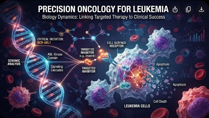
The article examines leukemia as an evolving population under targeted therapy, using chronic myeloid leukemia as the main example. BCR–ABL1 inhibition can produce deep responses, but every treatment changes the competitive environment among malignant clones. Quantitative transcript monitoring provides a continuous measure of disease burden, while deep sequencing can identify emerging resistance mechanisms before hematologic progression. A detected mutation is clinically meaningful only when its
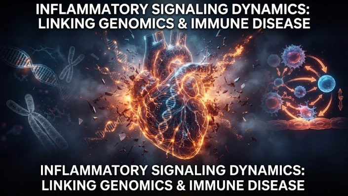
The article examines how cardiovascular genetic risk becomes biologically active through cell-specific inflammatory signaling. Because many vascular-risk variants lie in regulatory DNA rather than coding regions, their effects may appear only in particular endothelial, macrophage, smooth-muscle, or lymphocyte states. Single-cell studies of human atherosclerotic lesions and multiomic endothelial experiments are used to illustrate this context dependence. The article also discusses IL6-related sig
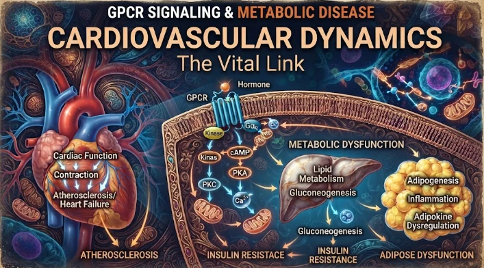
The article examines cardiovascular GPCR signaling as a dynamic response system whose abnormalities can remain hidden in resting measurements. Mechanical stretch, hormones, nutrients, sympathetic activity, and fluid balance can change the amplitude, duration, localization, and recovery of receptor signaling. Examples include mechanically activated angiotensin-receptor signaling, endosomal signaling, beta-arrestin-dependent trafficking, and biased glucagon-like peptide-one receptor responses. The