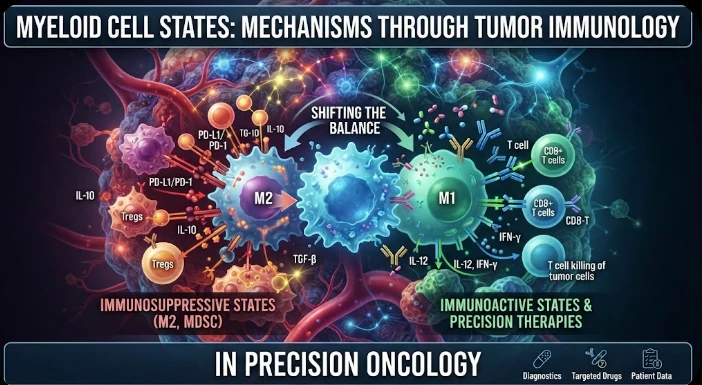
The article examines tumor-associated myeloid cells as diverse functional states rather than a single suppressive macrophage population. Pan-cancer single-cell studies reveal multiple macrophage and dendritic-cell programs with distinct developmental origins, spatial niches, and relationships to T cells, vasculature, fibrosis, and therapy. TREM2-positive macrophages are used as an example of a state that can be experimentally targeted to reshape the immune environment and improve checkpoint response in models, while cross-cancer variation cautions against one-marker rules. The article recommends longitudinal single-cell, chromatin, surface-protein, and spatial profiling to determine when myeloid states emerge under treatment. CRISPR perturbation, lineage analysis, coculture, organoids, and humanized models can test causal regulators and tumor–myeloid feedback loops. Clinical translation requires state-specific biomarkers, independent validation, safety assessment, and ecosystem-level monitoring. Precision oncology should therefore target reproducible myeloid dependencies without broadly compromising protective immune functions.
Previously, tumor associated myeloid cells have been classified as either suppressors (macrophages) or inflammatory (macrophages, monocytes and dendritic cells). This vocabulary is too coarse for precision oncology, as has been demonstrated with single cell technologies. There are numerous myeloid states present in human tumors that share some markers and have different developmental origins, but also have distinct characteristics of their relationships to T cells and vasculature, fibrosis, and responses to therapy. The main mechanistic question is what are the states that actively promote tumor progression or resistance, and what are those that just arise as a consequence of the local environment?
A single cell atlas of tumor infiltrating myeloid cells (TIMs) in over two hundred patients from fifteen different cancers showed that there is significant inter-tissue diversity. Macrophage and dendritic cells had common and tumor-specific characteristics. This information questions the assumption that there is a single marker that defines one suppressive population and its meaning in all cancer. Cell identity needs to be read in the context of lineage, tissue and transcripts.
One example that emerged was the TREM2 positive macrophages. Experimental studies revealed that targeting TREM2 was capable of reshaping the myeloid landscape in mouse models of tumors and improving the efficacy of PD-1 blockade. Selected macrophage sub-populations were depleted and immunostimulatory myeloid states enhanced by deletion or antibody targeting. The outcome showed causal evidence that altering a myeloid program could impact T cell response and not just correlate with tumor outcome.
However, TREM2 is not a pan macrophage marker. Subsequent single cell analyses identified TREM2 positive populations in relation to the FOLR2 positive and other states and the associations with prognosis were found to differ between cancer types. This is heterogenous and is important for target development. If there are insufficient details, a therapy targeting a broadly expressed myeloid marker might eliminate both protective and suppressive cells.
Longitudinal profiling can determine what state these occur under when treated. Biopsy (baseline) provides a determination of pre-treatment composition and early on treatment sampling can provide evidence of myeloid recruitment, differentiation or disappearance prior to radiographic response. Progression samples can be used to differentiate transient adaptation from persistent resistance. Paired samples are particularly useful as each patient acts as baseline for change.
Single cell RNA sequencing determines transcriptional state, and more layers brings more mechanism. Chromatin accessibility can indicate a regulatory program ready to be activated; surface proteomics can pinpoint targetable receptors; and spatial profiling can reveal the location of a state. A macrophage that surrounds T cells in an exhausted state has different functional implications than a similar transcriptional cell in the vicinity of necrosis/vasculature. Location can be a cell label, which becomes an interaction hypothesis.
In human tumors, lineage tracing is challenging and possible clues are provided by computational trajectories and somatic mutations. Macrophages that infiltrate a tumour can be induced to adopt different states by cytokines, hypoxia, lipid exposure and signals from the tumour. Matched blood and tumor populations can be used to measure likely transitions. Experimental models can then be used to determine if nominated signal induces differentiation towards the patient state.
CRISPR perturbation can be used to reveal regulators not markers. Myeloid cells can be genetically altered in a suppressive state to assess the effects of these disruption on differentiation, phagocytosis, cytokine production, and T cell activation. A causal regulator must be functionally changed upon perturbation. Pathway can be restored in rescue experiments and specificity can be confirmed. Testing of this type is vital before any cell marker is considered a therapeutic target.
Myeloid behavior is also influenced by the tumor cells. Macrophages can be instructed by cytokines, metabolites, extracellular nucleotides, lipids and products of dying cells. Myeloid cells in turn regulate angiogenesis, antigen presentation, ECM, and T cell access. It's a two-way street. The cancer cells in the tumor should thus be linked to myeloid cells, not the immune compartment itself.
These feedback loops can be identified by spatial multiomics. Communication pathways can be nominated by ligand/receptor expression and multiplex imaging can validate the finding of physically colocalized interacting populations. Coexpression does not provide evidence for signal transfer, so Computational Communication (CC) scores are hypotheses. Direction of function needs to be confirmed by perturbing the receptor, the ligand or the downstream pathway in co culture or tumour models.
Myeloid dependencies can be tested for in patient derived models to see if they survive outside the mouse system. Selected human interactions can be maintained in tumor organoids, with autologous immune cells and humanized models. These systems are capable of comparing the effects of checkpoint blockade alone with combinations of myeloid targeted therapies. Treatment can be molecularly profiled to determine if it is having an actual effect on the predicted state or just a nonspecific inflammatory effect.
Avoid one marker rules in clinical biomarker development. A combination of genes, proteins and spatial relationships can be more specific than TREM2 or one of the other receptors. The discovery signature can subsequently be simplified to a practical assay if reduced measurements can still be used to classify it. The state detection performance and association with treatment outcome requires external validation on independent cancer types.
The goals of causality and prediction should not be confused. Myeloid state can be a predictor of poor response as it is not a direct driver to resistance but rather aggressive tumor biology. On the other hand, a causal state may be either too scarce or too complex to be of prediction value. Each candidate should be considered for both functions (mechanistic necessity and clinical informativeness) to enable precision oncology.
Treatment combinations need safety consideration. Myeloid pathways play a role in infection defense, tissue repair and normal immune regulation. Widespread thinning may lead to toxicity or cause a lack of host protection. While state selective targeting or short treatment windows might lower risk, it will be necessary to ensure that the targeted program can only be administered within the tumor, and there is evidence that this is the case. Monitoring of pharmacodynamics should confirm selective remodeling and not systemic myeloid collapse.
The developmental origin can affect the function of the myeloid cells without influencing their actual state of transcription. Early life resident tissue macrophages can have similar phenotypes as monocyte derived macrophages recruited during tumor growth, but have different epigenetic history. Origin separation with single cell RNA is sometimes challenging. Models, lineage markers, and chromatin profiles can be used to test if two similar states follow different pathways and can influence the ease of reprogramming each state.
A moving therapeutic target is also provided by a myeloid plasticity. One program can be suppressed and another program fills in the niche. Design in combination should therefore not focus on the targeted cluster, but on the whole myeloid compartment. The success of the intervention could be assessed as ecosystem remodeling rather than only by the elimination of one population of cells that is positive for a given marker.
Imaging can be used in conjunction with biopsy to follow the myeloid rich areas over time. Selected immune targets can be estimated by emerging radiotracers/molecular imaging methods in whole body lesions and provide a different picture of the lesion heterogeneity than one biopsy. These tools must be highly specific as they may also become taken up in nonmalignant inflammation. Imaging can be used to aid in identifying lesions in which a myeloid state is dominant, when validated against paired tissue, and to direct sampling.
Stromal and vascular effects should also be explored in resistance studies. Myeloid cells can trigger angiogenic, matrix remodeling, and wound repair processes which are favorable to tumor growth, even in the absence of any apparent T cell suppression. A therapy that has the effect of enhancing T cell activation but also leaving a robust proangiogenic myeloid niche behind can generate partial benefit. In addition to lymphocyte assays, functional readouts should also contain vascular behavior, matrix state, phagocytosis and tumor cell growth.
Miriam Merad's research focus is on myeloid cell biology and human tumor immunity, a field revolutionized by single cell resolution. Today, precision oncology is shifting beyond macrophage counting to mapping their state, origin, location and function. The combination of multiomics and perturbation can identify which myeloid programs are "therapy-adaptive" and which are "therapy-causal". The useful target is a reproducible state dependency that can be modified without compromising the immunity.
CITE THIS ARTICLE
Tasmeem, S. (2025). Myeloid Cell States Mechanisms through Tumor Immunology in Precision Oncology. https://www.sadiaresearchhub.com/article/5f521d32-69e3-5b5b-a257-80cc57e4c85d
S. Tasmeem, “Myeloid Cell States Mechanisms through Tumor Immunology in Precision Oncology,” 2025. [Online]. Available: https://www.sadiaresearchhub.com/article/5f521d32-69e3-5b5b-a257-80cc57e4c85d
Tasmeem, S. (2025) ‘Myeloid Cell States Mechanisms through Tumor Immunology in Precision Oncology’. Available at: https://www.sadiaresearchhub.com/article/5f521d32-69e3-5b5b-a257-80cc57e4c85d
Tasmeem, Sadia. “Myeloid Cell States Mechanisms through Tumor Immunology in Precision Oncology.” 2025, https://www.sadiaresearchhub.com/article/5f521d32-69e3-5b5b-a257-80cc57e4c85d.
Sadia Tasmeem. “Myeloid Cell States Mechanisms through Tumor Immunology in Precision Oncology.” (2025). https://www.sadiaresearchhub.com/article/5f521d32-69e3-5b5b-a257-80cc57e4c85d.
Tasmeem S. Myeloid Cell States Mechanisms through Tumor Immunology in Precision Oncology. 2025. https://www.sadiaresearchhub.com/article/5f521d32-69e3-5b5b-a257-80cc57e4c85d
@misc{article_5f521d3269e35b5ba25780cc57e4c85d,
  title = {Myeloid Cell States Mechanisms through Tumor Immunology in Precision Oncology},
  author = {Sadia Tasmeem},
  year = {2025},
  url = {https://www.sadiaresearchhub.com/article/5f521d32-69e3-5b5b-a257-80cc57e4c85d},
}
TY  - GEN
AU  - Sadia Tasmeem
TI  - Myeloid Cell States Mechanisms through Tumor Immunology in Precision Oncology
PY  - 2025-11-16
UR  - https://www.sadiaresearchhub.com/article/5f521d32-69e3-5b5b-a257-80cc57e4c85d
ER  -
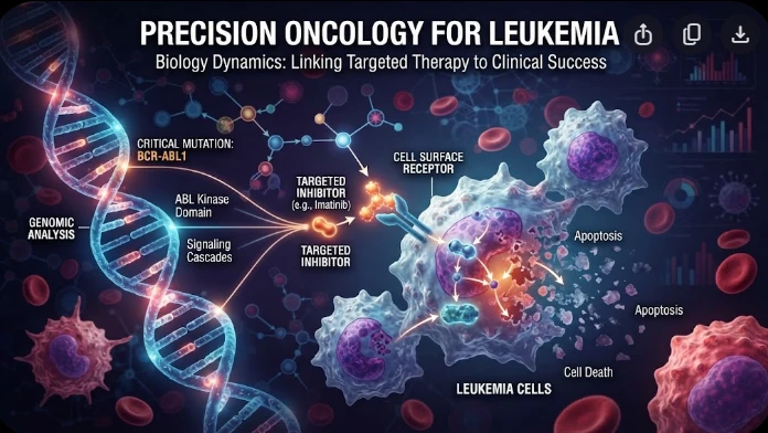
The article examines leukemia as an evolving population under targeted therapy, using chronic myeloid leukemia as the main example. BCR–ABL1 inhibition can produce deep responses, but every treatment changes the competitive environment among malignant clones. Quantitative transcript monitoring provides a continuous measure of disease burden, while deep sequencing can identify emerging resistance mechanisms before hematologic progression. A detected mutation is clinically meaningful only when its
The article examines how cardiovascular genetic risk becomes biologically active through cell-specific inflammatory signaling. Because many vascular-risk variants lie in regulatory DNA rather than coding regions, their effects may appear only in particular endothelial, macrophage, smooth-muscle, or lymphocyte states. Single-cell studies of human atherosclerotic lesions and multiomic endothelial experiments are used to illustrate this context dependence. The article also discusses IL6-related sig
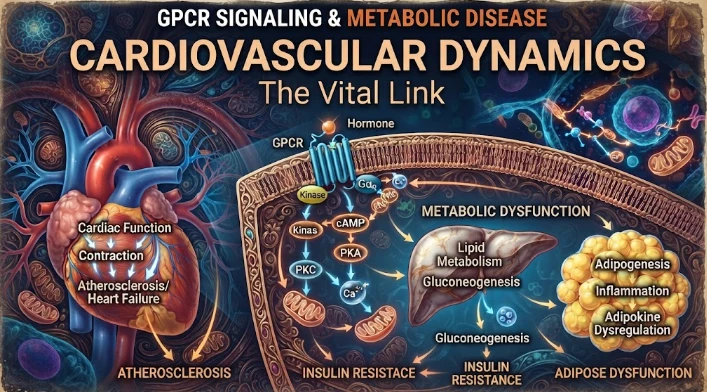
The article examines cardiovascular GPCR signaling as a dynamic response system whose abnormalities can remain hidden in resting measurements. Mechanical stretch, hormones, nutrients, sympathetic activity, and fluid balance can change the amplitude, duration, localization, and recovery of receptor signaling. Examples include mechanically activated angiotensin-receptor signaling, endosomal signaling, beta-arrestin-dependent trafficking, and biased glucagon-like peptide-one receptor responses. The