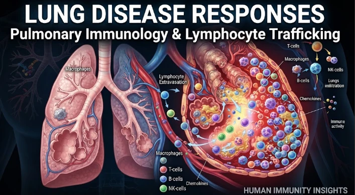
The article connects lung-disease outcomes with pulmonary immunology by focusing on how lymphocyte movement differs across infection, allergy, autoimmunity, and chronic inflammation. The lung is treated as a set of distinct immune compartments rather than a single space, with blood vessels, airways, interstitium, alveoli, and lymphoid structures providing different signals. Chemokines, receptor desensitization, histamine pathways, resident-memory populations, and tissue remodeling all influence whether recruited lymphocytes protect tissue or prolong damage. The article recommends combining blood, bronchoalveolar, sputum, tissue, imaging, single-cell, receptor-sequencing, and spatial data to identify whether disease reflects excessive recruitment, persistent residency, or failed regulation. Experimental receptor blockade and tissue models can test causality. Timing is especially important because a pathway can be beneficial early in infection and harmful later. The article ultimately proposes mechanism-based trafficking classifiers to guide stage-specific pulmonary immunotherapy while preserving protective immunity.
Lung disease can manifest with the same symptoms (although not always) from very different immune processes. Symptoms of cough, breathlessness, airflow limitation and radiographic inflammation may occur due to any of the following factors: infection, allergy, autoimmunity, environmental injury, and chronic remodeling. Pulmonary immunology then provides mechanistic resolution, that is, by asking which immune cells are present, what signals regulate their location, and what is the effect of these cells? protection of tissue, or propagation of damage? Lymphocyte trafficking is a big component of that solution because it links systemic immunity to local lung response.
The respiratory tract is not a single space, but multiple immune compartments. Different microenvironments of blood vessels, interstitium, airway epithelium, alveoli, draining lymph nodes and inducible lymphoid structures. It is therefore possible for the same lymphocyte to be present in a vascular margin, an epithelial lesion, or a long lived resident niche in the same tissue.
The antigens, cytokines, oxygenation, metabolites and stromal signals that will be encountered by the cell at each location are different.
A considerable part of this distribution is organized by chemokines. Other families of chemokines direct other subsets of lymphocytes to inflamed tissue, and interferon inducible ligands for CXCR3 actively attract activated T cells. Gradient strength and duration may affect the number of cells reaching an area and/or the distribution of cells. There is a counterweight to that, called receptor desensitization, which means that the receptors don't keep moving toward each inflammatory signal they come across during the immune response.
Interleukin sixteen is one such example of regulatory complexity as the CD4 signaling will affect the responsiveness to CXCR3. The cross talk alters the migration properties of activated T cells, even in the absence of loss of the receptor. Another pathway is via histamine H4 receptor signaling, which is involved in recruiting regulatory T cells to airway inflammation models. Pulmonary immune response is, however, generated in a cross-talked navigation system and not from individual chemokine modules.
In infection, the trafficking needs to be quick enough to stop pathogen replication but not cause excessive damage. Antigen specific T cells that invade the tissue after peak virus replication cannot help prevent tissue damage unless there is a direct antiviral benefit. Another pattern may be established by fungal or bacterial infection, where antigen is persistent, recruiting for a longer time. Immune traffic pattern is optimal depending on pathogen biology and stage.
The allergic lung disease is a different story. Exposure of environmental antigen may maintain recruitment of eosinophilic inflammation and mucus production and airway hyperreactivity-promoting helper T cell populations. The severity of the disease is the balance of entry, localization and suppression by regulatory lymphocytes. The therapeutic effect can occur with a shift in traffic and not just by decreasing overall cell numbers.
Structural change is added to the picture in chronic inflammatory lung diseases. The resulting damage to the epithelium, changes to the ECM, vascular remodeling and continued cytokine production can produce niches that continually attract or retain immune cells. These tissue microenvironments can perpetuate inflammation even if the initial cause of inflammation has diminished. Lymphocyte trafficking is used as a response to disease but also as a means to maintain disease.
It is further complicated by resident memory T cells. These cells stay in tissue following previous antigen exposure and are capable of responding quickly within the local area. This memory can help protect against repeat infection and an exaggerated resident response can play a role in chronic inflammatory pathology. Functional assays – not just cell counts or marker expression – are needed to distinguish protective residency from pathogenic persistence.
Immune movement can be reconstructed from human studies using bronchoalveolar lavage, blood, sputum, tissue biopsy, imaging and clinical physiology. Sequencing of the T cell TCRs can connect clonotypes between compartments. Single-cell transcriptomics can distinguish between activation and residency status. Spatial methods can identify if a clone is localized in the vicinity of infected epithelium, fibrotic tissue, blood vessels, immune aggregates etc. Then, repeated sampling can be used to determine if that population cleared up or did not.
Experimental support is required for causal interpretation. Tissue models could be used to evaluate the effect endothelial or epithelial signals have on migration. Blocking receptors can make a difference between the need and the lack of need for a chemokine axis. Recruitment and local proliferation and survival can be separated by performing perturbation experiments.
Animal models enable a controlled infection/allergen exposure and measurement of cell movement and lung function. Translation is optimal when there are similar trafficking signatures in human disease.
It is important to pay particular attention to variation within subgroups. Pulmonary immune responses are influenced by age, sex, smoking history, metabolic disease, immune suppression, previous infection, vaccinations, and drug exposure. Trafficking signatures that are seen in the recovery process in one group may be different in another. Mechanisms conserved across the cohorts can be distinguished from those host context-specific by multicohort analysis and prespecified subpopulation testing.
Clinical biomarkers should reflect mechanism, not inflammation. Airway chemokines, receptor state of lymphocytes, clonotype expansion, localization and functional measurements may allow for categorization of whether the disease is a consequence of ongoing recruitment, resident activation, or regulatory failure. It's a model that could help with more targeted immunotherapy. This is not something that can be achieved by measuring a single cytokine.
The timing of treatment is also crucial. Early inhibition of recruitment during infection may hinder clearance, and chronic inhibition of recruitment following pathogen control may diminish tissue damage. Regulatory trafficking may be desirable for allergic inflammation, but it may not be desirable if a potent antimicrobial effector function is needed. Trafficking-focused therapy will become the norm only when there are markers that can identify the transition from beneficial to damaging immunity, which is the phase that needs to be identified.
A variety of pulmonary diseases can also share the same chemokine and yield different cellular responses. CXCL10 may be recruited to participate in antiviral immunity, autoimmune inflammation, and chronic tissue damage, but with different recruited populations and context. The concentration of ligands, their state at the receptor, clonotype and tissue localization should therefore be integrated into a biomarker panel. This prevents using one inflammatory molecule as a disease-specific mechanism.
The study of recovery should be equal to that of peak disease. Resolution demands falling recruitment signals, removal/reprogramming of inflammatory cells, restoration of epithelial barrier function and survival of useful memory populations. Long-term follow-up, as in longitudinal cohorts that follow individuals after symptoms have improved, can establish predictors of lasting recovery from trafficking programs and of ongoing deficits. These data can provide information about therapeutic windows that are not captured by studies that follow patients through to hospital discharge or initial response.
Access by immune cells is also affected by mechanical forces. Changes in endothelial contact and tissue movement may result from changes in pulmonary blood flow, vascular pressure, edema, and altered matrix stiffness. Inflammation can then have a profound effect on trafficking by altering both the chemokine signaling pathways and the physical structure of the trafficking. If there is any context to be added, imaging and biomechanical measurements might help explain why a cell is found in one area of the lung and not another when molecular signals do not.
A meaningful trafficking classifier should be able to distinguish at least three states: deficient recruitment of protection, over-inflated entry of inflammation, and retention in tissue after the trigger is lowered. Every state has its own type of intervention. Immunity enhancement, selective blockade, and promotion of resolution/egress could help the first, second, and third, respectively. To distinguish these mechanisms and to change the movement of lymphocytes requires precision.
An additional source of variation may be added by the microbiome signals. Airway and gut microbes modulate the immune tone of the system and mucosal immune system prior to the arrival of lymphocytes at the lung. Therefore the baseline trafficking response may also be altered by the presence of antibiotics, infection history and chronic colonization. Cohort studies which document such exposures can help minimize unexplained heterogeneity and uncover host circumstances under which a protective pathway turns pathogenic.
This context enhances understanding.
Thus, the reactions of the human lung to disease can be considered as a movement of immunity in a dynamic tissue landscape. Pulmonary immunology provides understanding of the signals that cause that movement and spatial and longitudinal approaches describe its result. The question that is most informative is not whether lymphocytes are increased, but whether the right populations arrive at the right time and in the right compartment and depart when their mission is fulfilled. That framework connects trafficked biology to recovery, persistence, and immune mediated injury in a direct manner.
CITE THIS ARTICLE
Tasmeem, S. (2026). Lung Disease Responses through Pulmonary Immunology and Lymphocyte Trafficking in Human Immunity. https://www.sadiaresearchhub.com/article/bfbac970-9d68-5857-9b1d-c6d71464ea33
S. Tasmeem, “Lung Disease Responses through Pulmonary Immunology and Lymphocyte Trafficking in Human Immunity,” 2026. [Online]. Available: https://www.sadiaresearchhub.com/article/bfbac970-9d68-5857-9b1d-c6d71464ea33
Tasmeem, S. (2026) ‘Lung Disease Responses through Pulmonary Immunology and Lymphocyte Trafficking in Human Immunity’. Available at: https://www.sadiaresearchhub.com/article/bfbac970-9d68-5857-9b1d-c6d71464ea33
Tasmeem, Sadia. “Lung Disease Responses through Pulmonary Immunology and Lymphocyte Trafficking in Human Immunity.” 2026, https://www.sadiaresearchhub.com/article/bfbac970-9d68-5857-9b1d-c6d71464ea33.
Sadia Tasmeem. “Lung Disease Responses through Pulmonary Immunology and Lymphocyte Trafficking in Human Immunity.” (2026). https://www.sadiaresearchhub.com/article/bfbac970-9d68-5857-9b1d-c6d71464ea33.
Tasmeem S. Lung Disease Responses through Pulmonary Immunology and Lymphocyte Trafficking in Human Immunity. 2026. https://www.sadiaresearchhub.com/article/bfbac970-9d68-5857-9b1d-c6d71464ea33
@misc{article_bfbac9709d6858579b1dc6d71464ea33,
  title = {Lung Disease Responses through Pulmonary Immunology and Lymphocyte Trafficking in Human Immunity},
  author = {Sadia Tasmeem},
  year = {2026},
  url = {https://www.sadiaresearchhub.com/article/bfbac970-9d68-5857-9b1d-c6d71464ea33},
}
TY  - GEN
AU  - Sadia Tasmeem
TI  - Lung Disease Responses through Pulmonary Immunology and Lymphocyte Trafficking in Human Immunity
PY  - 2026-05-31
UR  - https://www.sadiaresearchhub.com/article/bfbac970-9d68-5857-9b1d-c6d71464ea33
ER  -
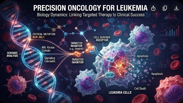
The article examines leukemia as an evolving population under targeted therapy, using chronic myeloid leukemia as the main example. BCR–ABL1 inhibition can produce deep responses, but every treatment changes the competitive environment among malignant clones. Quantitative transcript monitoring provides a continuous measure of disease burden, while deep sequencing can identify emerging resistance mechanisms before hematologic progression. A detected mutation is clinically meaningful only when its
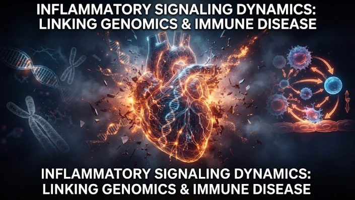
The article examines how cardiovascular genetic risk becomes biologically active through cell-specific inflammatory signaling. Because many vascular-risk variants lie in regulatory DNA rather than coding regions, their effects may appear only in particular endothelial, macrophage, smooth-muscle, or lymphocyte states. Single-cell studies of human atherosclerotic lesions and multiomic endothelial experiments are used to illustrate this context dependence. The article also discusses IL6-related sig
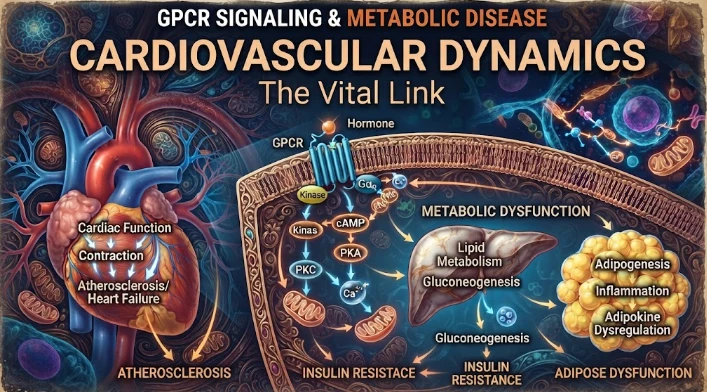
The article examines cardiovascular GPCR signaling as a dynamic response system whose abnormalities can remain hidden in resting measurements. Mechanical stretch, hormones, nutrients, sympathetic activity, and fluid balance can change the amplitude, duration, localization, and recovery of receptor signaling. Examples include mechanically activated angiotensin-receptor signaling, endosomal signaling, beta-arrestin-dependent trafficking, and biased glucagon-like peptide-one receptor responses. The