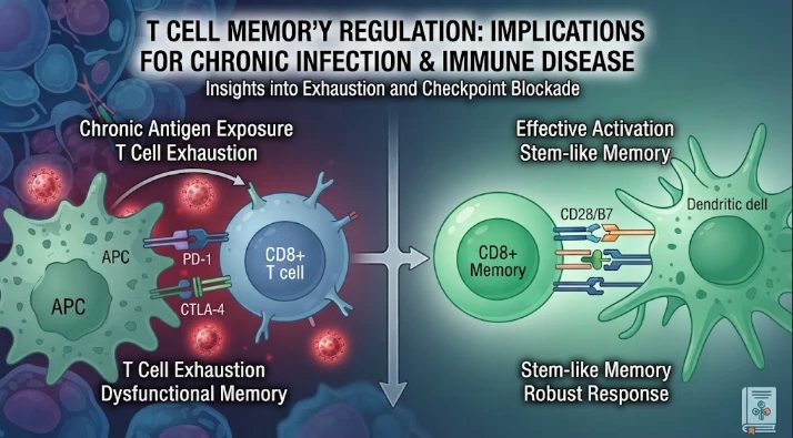
The article explores how chronic viral infection reshapes T-cell memory rather than simply reducing or preserving memory-cell numbers. Persistent antigen and inflammation can drive exhaustion, alter bystander memory cells, change proliferative capacity, cytokine production, metabolism, epigenetic state, and tissue distribution. Experimental chronic-infection models and human observations are used to show that antigen-specific and unrelated memory compartments can both be remodeled. The article argues that cross-sectional markers cannot distinguish durable memory from persistent dysfunctional cells. Longitudinal sampling should combine viral burden, single-cell transcriptomics, chromatin profiling, T-cell receptor sequencing, antigen-specific stimulation, proliferation, cytokine, and killing assays. Tissue-resident populations and metabolic reserve are also emphasized because blood measurements may miss important local states. Therapy and vaccination act as perturbations that can reveal reversible versus fixed dysfunction. Clinically useful memory signatures should predict rebound, vaccine response, durable suppression, or reinfection rather than merely describe cell phenotype.
The memory of T cells is typically thought of as the long-term memory of previous infections, but the nature of the memory of chronic viral infection is different. Repeated antigenic stimulation, chronic inflammation, changed tissue environments, and persistent antigen can change differentiation even after the acute response has contracted. The question of whether or not memory cells can be detected is not the central issue. For functional memory, proliferative capacity, cytokine production, tissue access, metabolic fitness, epigenetic state and a rapid response in the event of the return of antigen are all key factors.
Acute infection usually results in a rapid increase in virus specific T cells followed by a contraction until memory T cells are formed. Chronic infection means that there is a constant pressure. Antigen specific CD8 T cells become exhausted, over time losing function and changing their transcriptional programs, with progenitor-like populations maintaining function and facilitating renewal. Memory and exhaustion are thus different, but related, biological programs. Seeing all remaining virus-specific cells as identical can make it difficult to determine how they are being regulated to protect or to promote therapeutic response.
Long-lasting infection can even change the memory cells which don't recognize the chronic virus. In a chronic lymphocytic choriomeningitis virus model, it was found that the bystander memory CD8 T cells had an altered phenotype and function, and that the bystander memory CD8 T cells had decreased production of cytokines and lowered secondary expansion, as a consequence of interleukin six and STAT1 signaling. That discovery is an example of a systems effect, in that the immune environment is altered overall, rather than the antigen specific compartment alone. Therefore, a previous immune "memory" may be over-written by a subsequent persistent infection.
The second layer is added in human studies. Long-term differences in memory CD4 T cell programs have been observed after SARS-CoV-2 infection, with chromatin accessibility and proliferative capacity found to be different. The imprint of infection-associated inflammation was different to vaccine priming. This evidence provides support for a model that offers that the context of antigen exposure affects the quality of long term memory. While antigen identity is important, inflammatory states as a part of priming can also have a significant impact on the future response.
The transitions cannot be resolved by cross sectional immune profiling. If memory markers are present in a blood sample at one point in time, and there is no other time point, it does not indicate which of the above is going on in a population of cells. Viral load and treatment changes can be used to reconstruct trajectories by prospective sampling before, during, and after viral load or treatment changes. Single cell transcriptomics, functional assays, chromatin profiling and T cell receptor sequencing can reveal lineage relationships, and assess the actual recall capacity.
Antigen specific assays are of paramount importance. Each of the assays (peptide stimulation, tetramer based detection, cytokine measurements, proliferation assays, and killing assays) ask a different question. A cell may have a memory phenotype, but not be able to proliferate well; may also have a memory phenotype, but not make any cytokines with strong cytotoxic activity. Incorporating functional readouts with molecular profiling prevents the false impression of marker expression is protection. An antigen specific measurement along the axes can also be used to differentiate persistent stimulation from real memory after viral control.
The interpretation of tissue context adds to the complexity. The blood is available but is typically only 10% of the relevant T-cell populations. Tissue resident memory cells in mucosal sites, liver, lymphoid organs or other compartment of infection can have different rules compared to circulating cells. Viral reservoirs can sustain local exposures to antigen (in the absence of viral plasma levels). If ethically and clinically appropriate, tissue sampling can identify if there is apparent systemic control in the presence of local immune pressure.
Therapy is a natural perturbation. The sharp reduction in antigen achieved with antiviral therapy has the potential to allow the identification of which T cell states are maintained over time without continued stimulation and which recover function after viral suppression. In selected research situations, immune checkpoint inhibition can be used to assess whether or not inhibitory pathways are keeping the immune system dysfunctional and even certain, though not full, reversions can be accomplished. Vaccination of chronically infected individuals can test the recall function from an alternate perspective. The best designs are those that correlate intervention effects, antigen burden, cell state and functional response over time and not just two distinct groups at once.
Confounding has to be addressed for causal interpretation. Exposure to treatment, antigen burden, and tissue damage may all occur at the same time in severe infection to create stronger inflammation. Each factor can influence the T cell memory.
In the case of combined exposures, statistical adjustment will lessen the bias but will not remove it completely. Experimental models are still important for controlled perturbation, and human longitudinal cohort studies provide relevance over actual disease history. There is more concordance between the two levels of data than there is within each level.
The emphasis of clinical translation should be placed on functions that are relevant to control. Useful memory signatures would include predicting: rebound following treatment interruption; response to therapeutic vaccination; durable viral suppression; or susceptibility to reinfection. This signature requires external validation based on age, sex, previous exposure, viral genotype, presence of comorbidities and treatment history. Immune memory is not an inherent characteristic of the named subset of cells. It is an active capacity which is a reflection of the priming that has taken place and the ongoing biological pressure.
Under chronic stress, memory may stay intact or fail depending on the cellular processes. Flexible access to glucose, amino acids, mitochondrial respiration and lipid metabolism is essential for the rapid responding T cells, and the persistence of antigen can modify those programs. Metabolic measurements can then be used as an addition to transcriptional state. Cells with different reserve can be identified as phenotypically similar cells using mitochondrial membrane potential, respiratory capacity, expression of nutrient transporters, and metabolite profiling. Post-nutrient/oxygen stress functional recall is particularly useful as infected tissues may place additional stress on the tissues that is different from standard culture media.
Another dimension is provided by the clonal history. New populations of memory T cells following viral suppression may be evidence for a clonal expansion from existing memory cells during high antigen loads, or because of the recruitment of rare clones. Tracing the steps of clonotype with single cell state allows for reconstruction of lineage pathways. Different mechanistic meaning is given to a clone that emerges from a progenitor such as a state to an effective memory compared with a new clone emerging after antigen decline. Repeated sampling can differentiate persistence, replacement and expansion, and identify these as different, not all as frequency changes.
Clinical endpoints should be selected based on the biology. Each of the four functions tested (durable suppression, breakthrough infection, viral rebound, response to booster vaccination, and recovery after immune perturbation) tested different types of memory functions. No single biomarker that is predictive of one endpoint can be used to predict another. A memory signature may not become clinically actionable until the following criteria are met: Prespecified outcomes, independent replication and consistent antigen specific assays. The best signature would not only indicate the current state of immunity but also the potential for future immunity, so as to determine if there is enough cellular reserve remaining to mount an effective recall response after chronic exposure.
Another component that influences memory is the ability to coordinate with other compartments of the immune system. Dendritic cells regulate antigen presentation and the context of cytokines, B cells provide antibodies and antigen presentation, and innate cells provide inflammatory tone. Chronic infection can affect both partners at the same time. Systems immunology can thus determine if the failure of T cells is an intrinsic program or abnormal supporting environment or both. A breakdown of the relationship can be identified and then mechanistic interventions can be more precise.
Alternatively, functional reserve can be measured following a fixed period of rest. The recovery of proliferation and cytokine production following antigen reduction would be a sign of reversible suppression, or a more stable memory defect. The distinction can inform the type of therapy to be used: cut away from the antigenic pressure or re-program them directly.
Rafi Ahmed's research focus has been to bring the concepts of T cell memory, chronic infection and vaccination into a single mechanistic framework. This view emphasises the potential importance of a persistent virus as a long-term determinant of immune state, not as a sustained acute infection. Distinctive differentiation of durable protection from persistent, but less strong, cellular presence is possible via prospective single cell profiling, antigen specific functional testing, and clinical linkage. The important take-home message is not that there is a lot of memory marker present, but that a T-cell population is able to maintain its identity, withstand chronic stress, and respond appropriately when immune control is called upon.
CITE THIS ARTICLE
Tasmeem, S. (2026). T Cell Memory Regulation through Chronic Viral Infection in Immune Disease. https://www.sadiaresearchhub.com/article/6583aba8-5f76-5829-8226-da564075a049
S. Tasmeem, “T Cell Memory Regulation through Chronic Viral Infection in Immune Disease,” 2026. [Online]. Available: https://www.sadiaresearchhub.com/article/6583aba8-5f76-5829-8226-da564075a049
Tasmeem, S. (2026) ‘T Cell Memory Regulation through Chronic Viral Infection in Immune Disease’. Available at: https://www.sadiaresearchhub.com/article/6583aba8-5f76-5829-8226-da564075a049
Tasmeem, Sadia. “T Cell Memory Regulation through Chronic Viral Infection in Immune Disease.” 2026, https://www.sadiaresearchhub.com/article/6583aba8-5f76-5829-8226-da564075a049.
Sadia Tasmeem. “T Cell Memory Regulation through Chronic Viral Infection in Immune Disease.” (2026). https://www.sadiaresearchhub.com/article/6583aba8-5f76-5829-8226-da564075a049.
Tasmeem S. T Cell Memory Regulation through Chronic Viral Infection in Immune Disease. 2026. https://www.sadiaresearchhub.com/article/6583aba8-5f76-5829-8226-da564075a049
@misc{article_6583aba85f7658298226da564075a049,
  title = {T Cell Memory Regulation through Chronic Viral Infection in Immune Disease},
  author = {Sadia Tasmeem},
  year = {2026},
  url = {https://www.sadiaresearchhub.com/article/6583aba8-5f76-5829-8226-da564075a049},
}
TY  - GEN
AU  - Sadia Tasmeem
TI  - T Cell Memory Regulation through Chronic Viral Infection in Immune Disease
PY  - 2026-02-08
UR  - https://www.sadiaresearchhub.com/article/6583aba8-5f76-5829-8226-da564075a049
ER  -
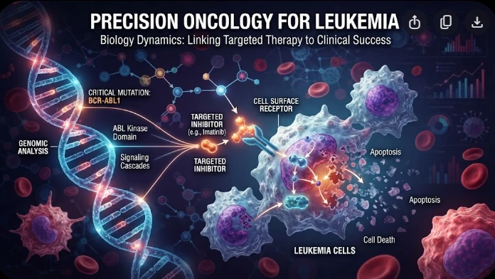
The article examines leukemia as an evolving population under targeted therapy, using chronic myeloid leukemia as the main example. BCR–ABL1 inhibition can produce deep responses, but every treatment changes the competitive environment among malignant clones. Quantitative transcript monitoring provides a continuous measure of disease burden, while deep sequencing can identify emerging resistance mechanisms before hematologic progression. A detected mutation is clinically meaningful only when its
The article examines how cardiovascular genetic risk becomes biologically active through cell-specific inflammatory signaling. Because many vascular-risk variants lie in regulatory DNA rather than coding regions, their effects may appear only in particular endothelial, macrophage, smooth-muscle, or lymphocyte states. Single-cell studies of human atherosclerotic lesions and multiomic endothelial experiments are used to illustrate this context dependence. The article also discusses IL6-related sig
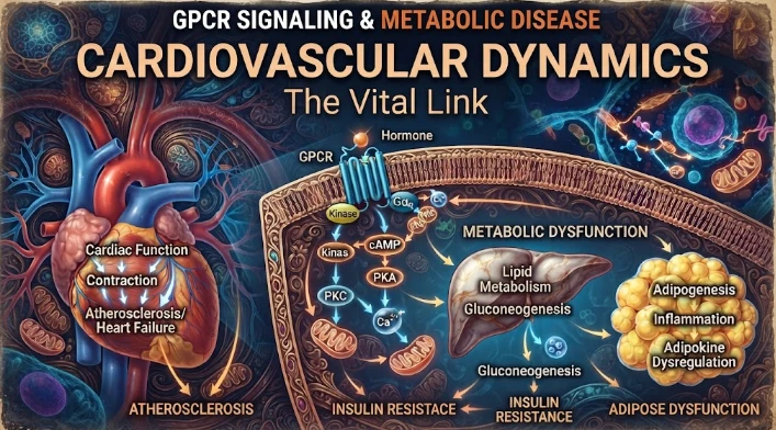
The article examines cardiovascular GPCR signaling as a dynamic response system whose abnormalities can remain hidden in resting measurements. Mechanical stretch, hormones, nutrients, sympathetic activity, and fluid balance can change the amplitude, duration, localization, and recovery of receptor signaling. Examples include mechanically activated angiotensin-receptor signaling, endosomal signaling, beta-arrestin-dependent trafficking, and biased glucagon-like peptide-one receptor responses. The