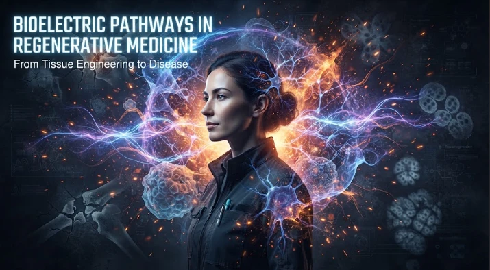
The article examines tissue regeneration as a process in which bioelectric changes can occur immediately after injury and influence later genetic repair programs. Damage alters membrane voltage, ion currents, gap-junction communication, calcium entry, second messengers, and transcriptional regulators. These early physiological signals can shape how stem cells proliferate, migrate, differentiate, and restore tissue structure. Proteomics is emphasized because channel abundance, trafficking, phosphorylation, and membrane localization can change faster than RNA levels. Longitudinal molecular measurements can identify which events precede successful healing and which accompany chronic injury. Human skin and epithelial wound models are proposed as accessible systems for testing whether early voltage patterns predict re-epithelialization, inflammation, or scarring. Functional rescue should demonstrate organized tissue repair, not merely increased proliferation. Local delivery approaches may reduce systemic electrical risks. The central argument is that regeneration is better understood by integrating fast bioelectric signals with slower transcriptional and structural repair mechanisms.
It is generally believed that regeneration is a series of molecular events: injury signals trigger the expression of genes, stem or progenitor cells divide, new tissue forms, and the anatomy is restored. Bioelectric research provides a very rapid physiological layer that starts essentially as soon as damage occurs. Membrane voltage and ion currents, as well as gap junction communication may shift prior to the onset of larger transcriptional programs, providing an early signal that will affect the way in which later regeneration proceeds.
The important variable is resting membrane potential. The ion channels, pumps and transporters in cells create different voltage states. Damage to tissue causes a change in the ionic environment and in the properties of the membranes. Those changes can affect the entry of calcium, the release of neurotransmitters, second messenger pathways, and transcription factors, and thus the regulation of genes, which is the connection of an electrical event with the regulation of genes.
The planarian head is regenerated in the following sequence. Altering the membrane potential by pharmacologic or genetic manipulation can affect the anterior identity. Early electrical perturbation is done prior to the normal head tail distinction set up by downstream patterning genes. The tissue as such is thus seen to rely on voltage as one of the decision factors, interpreting the position of the wound.
Timing is critical. Even after the physiological conditions become normal, a small disturbance in the first few hours following the amputation may result in a permanent anatomical effect. The experiments suggest that bioelectric signaling is a switch or a state selector rather than a daily force that physically moulds tissue.
Gap junctions pass the signal through cells. These channels give ions and small molecules free access between neighbors, creating a network of membranes within a tissue. Regenerative polarity is effected by altering the ability of gap junctions to communicate with one another, demonstrating the need for coordination in order to obtain anatomical information.
One pathway is voltage to molecular response via calcium. Neuromuscular function and patterning can be affected by voltage gated calcium channels in planaria. Transitions in calcium entry will have an effect on the regenerative outcome, suggesting that the change in bioelectric state results in a change of intracellular signals that affect gene expression and cell behavior.
Another downstream mechanism occurs through apoptosis/remodeling. In order to be able to regenerate correctly, new cells are not only generated but old tissue must be removed and replaced in a new location. Disruption of bioelectricity can change the number or distribution of apoptosis, and lead to abnormal organ scaling. The voltage may therefore play a role in the editing phase which brings the regenerating structure closer to the right proportion.
To determine the molecular components of these circuits, long read sequencing and transcriptomics can be useful. Often, you will find that there are multiple isoforms of a given ion channel gene: these may vary between tissues and/or developmental stages. Long reads are able to resolve transcript assembly problems caused by short sequencing reads. The expression data can then be used to highlight the channel variants that occur within the critical period of regeneration.
The functional layer is added by proteomics since the abundance and modification of channels are not necessarily deduced from the amount of RNA. Membrane proteins can be degraded, trafficked or phosphorylated quickly after injury. The detection of channel proteins, pumps and signaling intermediates over time can link the electrical phenotype to the molecular machines that generate it.
Models of independent diseases are required for translation. Discoveries made in planaria tissue must be tested in engineered human tissue, or in amphibian or mammalian tissue, in order to assess its effect on human anatomy and immune responses. Conservation of a voltage dependent pathway across models would lend support to the idea that this is a general mechanism.
The information could be applied to regenerative medicine in several ways in the future. Tissue voltage can be changed by electrical stimulation, an ion channel drug or a targeted gene control or biomaterial electrodes. Short and local interventions that would get a wound to a regenerative state without chronic manipulation would be most attractive. It takes knowledge of the voltage pattern that is instructive and when the tissue is ready to respond in its newly created state.
Another cautionary example is cancer biology. Without pattern control, there are risks associated with promoting proliferation and migration of abnormal membrane potential. The coordinated anatomy is needed for regeneration, rather than just increased cell division. If bioelectric therapy were to be used, it would require endpoints of structure, function and tumor risk.
The early "wound" signals should be distinguished from later "maintenance" signals by experiment. A channel blocker can be given only in the first few hours to test initiation and a later treatment can test if the same channel is required for maintaining the pattern. The use of temporal restriction decreases the probability that a general drug effect on growth is misinterpreted as a polarity mechanism.
Imaging can be complemented by electrophysiology. Selected cells or tissues can be directly recorded for validation of voltage estimates by reporter. There are several factors that can impact the performance of reporters, such as dye loading, membrane permeability, or optical set-up. Consistency of results from independent measurement techniques supports the conclusion that a true bioelectric change was the precursor to regeneration.
Multiple samples can be taken downstream of the electrical perturbation. Immediate early genes, patterning genes and differentiation markers are not expected to happen simultaneously. A time ordered molecular cascade can be used to demonstrate the transition of a short voltage event to a days-long anatomical program.
As well as expression, protein localization can be as important. After injury, ion channels can be translocated into or out of the membrane, but there is not a significant change in the transcription of the channels. This fast regulation can be captured in the proteomics of membrane fractions or imaging studies of tagged channels. These protein-level measurements are most useful when used in conjunction with long read transcriptomics.
Independent replication between laboratories is particularly important as the regeneration experiments may rely on species, temperature, feeding state and amputation position. A well-defined protocol and common set of reference conditions can distinguish between a robust and sensitive bioelectric mechanism.
Human wound models can be used to evaluate the ability of early voltage changes to predict healing quality. Skin or epithelial injuries are easy to measure and re-sample using noninvasive electrical measurements. Leveraging conserved features that correlate with voltage reepithelialization, inflammation, and scar formation will provide insight into mechanistic follow up.
Drug specificity should be checked, as drugs which act on ion channels have several targets. An effect observed in the channel can be distinguished from the voltage dependent effect by genetic perturbation and by an unrelated drug that causes the same voltage change. The evidence is especially strong with convergent rescue.
Vascular and immune coordination is required for tissue repair as well. Macrophages, endothelial growth and nerve regrowth can be influenced by electric signals. Multi-cell models can simulate the direct or indirect effect of bioelectric manipulation on progenitor cells.
An anatomical state that is desired before intervention is a characteristic of a translational program. The safer option to regenerative biology is when the successful result is the restoration of a measured pattern rather than continual growth stimulation.
Local interventions can be made, and repeated measures of repair can be taken, in tissues that are readily accessible, including skin, cornea, and peripheral nerve (which are all used in mammalian translation). These models can be used to assess if voltage guided repair can functionally improve without systemic exposure. A bounded tissue is a better source of translationally relevant evidence than extrapolating directly from planaria whole body regeneration.
That progression might simulate a translation from the physiology of model organisms to bounded human tissue repair – but not quite stretch the regenerative potential.
As a researcher with an interest in bioelectric signaling and tissue regeneration, Michael Levin underscores the importance of studying regenerative mechanisms at different scales. Early ion current can change calcium, gene expression, apoptosis and finally whole body pattern. Experiments with voltage manipulation and downstream molecular changes with a rescue by an independent pathway are the strongest evidence. Bioelectric signaling is most relevant to human disease when it can be linked, in a reproducible mechanistic pathway, from membrane physiology to functional tissue repair.
Systemic electrophysiological risk can be reduced, as well. with local delivery. A scaffold, topical or device mediated intervention can expose the repair site and minimize heart and nervous system impact.
Delivery matters.
CITE THIS ARTICLE
Tasneem, S. (2025). Tissue Regeneration Mechanisms Linking Bioelectric Signaling to Human Disease. https://www.sadiaresearchhub.com/article/8c5560b9-f2a3-53c3-86e6-4d154813af7a
S. Tasneem, “Tissue Regeneration Mechanisms Linking Bioelectric Signaling to Human Disease,” 2025. [Online]. Available: https://www.sadiaresearchhub.com/article/8c5560b9-f2a3-53c3-86e6-4d154813af7a
Tasneem, S. (2025) ‘Tissue Regeneration Mechanisms Linking Bioelectric Signaling to Human Disease’. Available at: https://www.sadiaresearchhub.com/article/8c5560b9-f2a3-53c3-86e6-4d154813af7a
Tasneem, Sadia. “Tissue Regeneration Mechanisms Linking Bioelectric Signaling to Human Disease.” 2025, https://www.sadiaresearchhub.com/article/8c5560b9-f2a3-53c3-86e6-4d154813af7a.
Sadia Tasneem. “Tissue Regeneration Mechanisms Linking Bioelectric Signaling to Human Disease.” (2025). https://www.sadiaresearchhub.com/article/8c5560b9-f2a3-53c3-86e6-4d154813af7a.
Tasneem S. Tissue Regeneration Mechanisms Linking Bioelectric Signaling to Human Disease. 2025. https://www.sadiaresearchhub.com/article/8c5560b9-f2a3-53c3-86e6-4d154813af7a
@misc{article_8c5560b9f2a353c386e64d154813af7a,
  title = {Tissue Regeneration Mechanisms Linking Bioelectric Signaling to Human Disease},
  author = {Sadia Tasneem},
  year = {2025},
  url = {https://www.sadiaresearchhub.com/article/8c5560b9-f2a3-53c3-86e6-4d154813af7a},
}
TY  - GEN
AU  - Sadia Tasneem
TI  - Tissue Regeneration Mechanisms Linking Bioelectric Signaling to Human Disease
PY  - 2025-03-02
UR  - https://www.sadiaresearchhub.com/article/8c5560b9-f2a3-53c3-86e6-4d154813af7a
ER  -
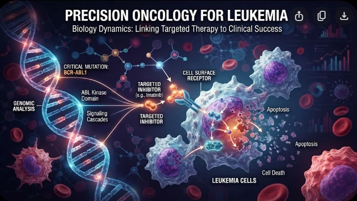
The article examines leukemia as an evolving population under targeted therapy, using chronic myeloid leukemia as the main example. BCR–ABL1 inhibition can produce deep responses, but every treatment changes the competitive environment among malignant clones. Quantitative transcript monitoring provides a continuous measure of disease burden, while deep sequencing can identify emerging resistance mechanisms before hematologic progression. A detected mutation is clinically meaningful only when its
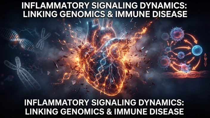
The article examines how cardiovascular genetic risk becomes biologically active through cell-specific inflammatory signaling. Because many vascular-risk variants lie in regulatory DNA rather than coding regions, their effects may appear only in particular endothelial, macrophage, smooth-muscle, or lymphocyte states. Single-cell studies of human atherosclerotic lesions and multiomic endothelial experiments are used to illustrate this context dependence. The article also discusses IL6-related sig
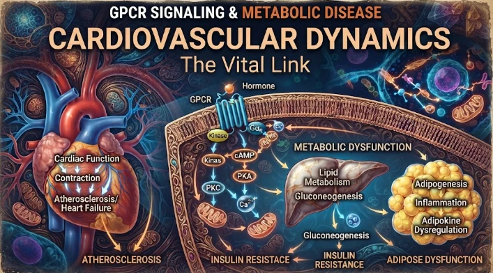
The article examines cardiovascular GPCR signaling as a dynamic response system whose abnormalities can remain hidden in resting measurements. Mechanical stretch, hormones, nutrients, sympathetic activity, and fluid balance can change the amplitude, duration, localization, and recovery of receptor signaling. Examples include mechanically activated angiotensin-receptor signaling, endosomal signaling, beta-arrestin-dependent trafficking, and biased glucagon-like peptide-one receptor responses. The