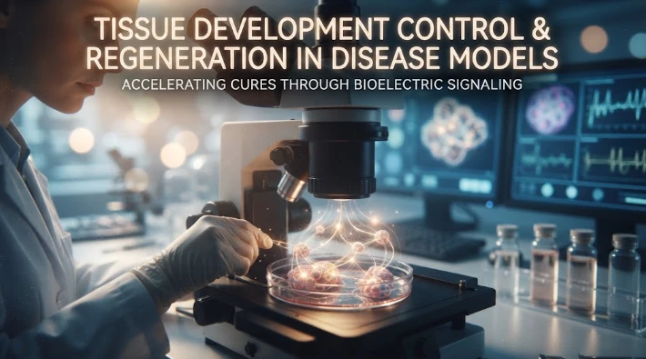
The article connects developmental biology and tissue regeneration through bioelectric signaling. Ion channels and pumps establish membrane-voltage differences, while gap junctions allow those signals to form multicellular patterns with boundaries and gradients. These electrical states can influence gene expression, cell identity, migration, and anatomical organization, providing physiological positional information alongside morphogens and genetic regulation. Disease models can reveal how altered channel or transporter function changes tissue-level voltage patterns and produces developmental, regenerative, or cancer-related phenotypes. The article recommends integrating bioelectric measurements with transcriptomics, chromatin profiling, imaging, genetic perturbation, and computational models that include voltage and biochemical gradients. Functional rescue should assess anatomy and organ performance because restored structure can still lack proper nerves, muscle, or vascular function. The overall message is that bioelectric physiology can help explain how tissues coordinate form, but translational claims require causal perturbation, reproducibility, and evidence that restored patterning also restores function.
Developmental biology is the study of how, out of the same genome, cells build different tissues in the right places. When wounding occurs, regeneration asks the question. Both problems are connected by using bioelectric signaling in that patterns of membrane voltage can give positional and state information across groups of cells. By altering these patterns with disease models, the roles of anatomy in physiology and how it can be controlled by physiological networks in concert with genetic and biochemical pathways is revealed.
The voltage difference across cell membranes is produced by the channels and pumps that transport ions. Adjacent cells are joined by gap junctions that enable electrical signals to spread to larger tissue areas. These circuits can give rise to stable patterns, with boundaries and gradients. These patterns may have a role in the development or regeneration of tissue in which they occur, by coordinating which genes are activated in what region.
The planaria test is particularly strong as a small piece of the body can regenerate the entire missing body. The effects of experiments that alter proton pumps, potassium channels, calcium channels, or gap junction communication can alter head tail identity, organ size, and other anatomical outcomes. The DNA sequence does not change, thus the modified form has to be the result of different interpretations of physiological information.
The most prominent aspect is multistability. Multiple stable bioelectric patterns can exist within the same Genome of a tissue. If the system is put into an abnormal head tail state by temporary manipulation, it can be kept in this state through the subsequent regens afterwards. This looks something more like a memory stored in a network state that doesn't look like a new mutation.
That concept can be formalized with the help of computational models. The multiple stable solutions of the pattern at the tissue-level can be produced by the cells with voltage dependent channels and gap junction coupling.The cells with the voltage dependent channels and gap junction coupling can generate the tissue level patterns with multiple stable solutions. Moving from one solution to another moves the network from one channel activity/connectivity to another. These models can make predictions on which perturbations should affect anatomy and when a state can be reset.
Gene regulation is still very much coupled. Alterations of positional genes may be preceded by early changes in voltage following amputation. There are pathways between membrane potential and transcription including calcium signaling and other second messengers. The electrical network thus serves as a "control" layer, in which it can skewer downstream genetic programmes towards one anatomical outcome.
Genes that are responsible for building up this layer can be identified using network based functional genomics. CRISPR perturbation can be used to test ion channels, pumps, gap junction proteins, and voltage sensitive signaling molecules. If multiple perturbations are combined, redundancy can be found — multiple channels can have the same voltage state. Network compensation can not be captured by single gene knockout.
Causal interpretation is enhanced by isogenic models. Genetically matched channel control cells or organisms can be used to compare with channel mutation cells or organisms. The restoration of the gene or correction of membrane voltage by another mechanism may be tested. Normalization of morphology in the face of the initial gene defect makes voltage more credible as causal variable.
Diseases don't just revolve around regeneration. Altered cellular physiology is involved in channelopathies, developmental malformations, cancer and wound healing. The disease action of a mutation in an ion transporter may be due not only to its actions in excitable cells but also to its actions on tissue level voltage patterns. Thus, a bioelectric approach can reveal phenotypes not known by conventional electrophysiology.
Appropriate scaling is required for human relevance. There are differences in the body plan of a planarian and a human and also in the regenerative capabilities. The real question is if mammalian tissues employ instructive voltage states during repair and development, too. Conserved mechanisms can be tested in organoids, embryos, wound models, and engineered tissues without taking them for granted as therapeutic equivalents.
Growth should NOT be the only functional endpoint included in a functional endpoint. Disorganized tissue can be a result of a treatment that encourages the growth of tissue. The correct polarity, size, connectivity, vascularization, and mechanical function are needed for regenerative success. In this context, the evaluation of bioelectric interventions should be made based on the restoration of the anatomical and physiological aspects, and not just based on the number of cells.
There are significant safety concerns. Ion channel modulation can influence the cardiac rhythm, neuronal excitability and smooth muscle. The pathways that regulate development can also be involved in cancer pathways. Local delivery, transient intervention and tissue specific targets are more feasible than systemic voltage manipulation.
There are windows of competence in developmental timing. Once tissues are mature, a voltage pattern which alters the anatomy would have little effect. Knowing these windows can provide insight into when downstream gene networks are open. Disease models equipped with developmental mutations can then be used to determine if a restoration of the correct electrical state within the limits of the window restores anatomy.
Left-right patterning and organ asymmetry may also engage in bioelectric network interactions. Differences that arise early in life can be magnified by later expression of genes. In selected models, it is possible to identify physiological asymmetry prior to molecular asymmetry by measuring bilateral voltage and channel activity.
A controlled test of pattern rules is possible using synthetic biology. Defined ion channel, gap-junction, and voltage sensitive transcriptional circuits can be provided to engineered cell collectives. One question for researchers is whether electrical states can be designed that generate predictable growth boundaries. These decrease biological complexity and provide tests of computational principles derived from animals.
A causal model should forecast disruption and rescue. If the second intervention is not specified in the model, then the intervention that results in an unusual body plan should be altered to restore the expected voltage state and body plan. If one perturbation can produce two phenotypes, it will be better evidence than if it could produce one.
Another criterion for clinical relevance is whether it is compatible with current repair methods. Bioelectric cues are potentially interacting with stem cell transplantation, biomaterials, growth factors, and rehabilitation. The electrical intervention may not be able to regenerate tissue on its own but rather guide implanted cells and/or enhance integration. That more realistic role can be tested by using combination studies.
Computational models can incorporate both bioelectric state and morphogen gradients. Voltage may alter the activity of the transport or receptor, and morphogens may affect channel expression. It is sometimes possible to determine why genetic and electrical perturbations can have similar anatomical phenotypes by testing both systems at the same time.
Patient-derived organoids with variants in gap junctions or ion channels can be used in disease modelling in development. Applying the variant to correct electrical organization and tissue pattern could help determine if they return to normal together. Such models make a more direct link between human genotype and a physiological mechanism than cross species analogy.
It's essential that it is stable in the long term. A form established at one time that results in an unstable network may be ineffective when damage or growth occurs later. Rechallenge experiments can be used to find out if the corrected pattern is robust.
Ethical translation must ensure that the body plan editing of the human body is not already possible with bioelectric control. Currently, there is more evidence in the experimental models. The next step that is scientifically valuable is the identification of conserved repair signals with bounded, localized effects which could be tested safely in mammalian tissue.
Developmental models should also be used to determine if it is possible to correct the electrical system after a genetic defect has already modified anatomy. The therapeutic questions of rescue before the formation of the pattern and rescue after malformation are different. The latter is more challenging, but more applicable to existing disease. That is why it is important to separate these windows, so that early developmental rescue does not appear to be a demonstration of reversing structural abnormalities that have already matured.
This control blinds bioelectric developmental research to testable medical issues instead of speculating with anatomical control.
The work of Michael Levin in bioelectric signaling and developmental biology provides a model for tissue form that is maintained by the interplay of genetic and physiological networks. Disease-models demonstrate that genome changes are not required to redirect anatomy through changing the electrical state. Network based functional genomics can help determine which channels establish these states and which downstream genes interpret the states. Recognizing the possibility of architecture to open up regenerative medicine not only to providing cells and growth factors, but also to directing the information to cells that determines the architecture of the structure they create.
If feasible, behavior or organ function should be included in functional rescue. There may be incomplete patterning even with correct anatomy if there is a failure of physiology in the nerves, muscles, or vascular connections.
The final test is physiology.
CITE THIS ARTICLE
Tasneem, S. (2025). Tissue Development Control through Bioelectric Signaling and Tissue Regeneration in Disease Models. https://www.sadiaresearchhub.com/article/bee7dcc0-36e9-5ad7-b689-7c4333f96454
S. Tasneem, “Tissue Development Control through Bioelectric Signaling and Tissue Regeneration in Disease Models,” 2025. [Online]. Available: https://www.sadiaresearchhub.com/article/bee7dcc0-36e9-5ad7-b689-7c4333f96454
Tasneem, S. (2025) ‘Tissue Development Control through Bioelectric Signaling and Tissue Regeneration in Disease Models’. Available at: https://www.sadiaresearchhub.com/article/bee7dcc0-36e9-5ad7-b689-7c4333f96454
Tasneem, Sadia. “Tissue Development Control through Bioelectric Signaling and Tissue Regeneration in Disease Models.” 2025, https://www.sadiaresearchhub.com/article/bee7dcc0-36e9-5ad7-b689-7c4333f96454.
Sadia Tasneem. “Tissue Development Control through Bioelectric Signaling and Tissue Regeneration in Disease Models.” (2025). https://www.sadiaresearchhub.com/article/bee7dcc0-36e9-5ad7-b689-7c4333f96454.
Tasneem S. Tissue Development Control through Bioelectric Signaling and Tissue Regeneration in Disease Models. 2025. https://www.sadiaresearchhub.com/article/bee7dcc0-36e9-5ad7-b689-7c4333f96454
@misc{article_bee7dcc036e95ad7b6897c4333f96454,
  title = {Tissue Development Control through Bioelectric Signaling and Tissue Regeneration in Disease Models},
  author = {Sadia Tasneem},
  year = {2025},
  url = {https://www.sadiaresearchhub.com/article/bee7dcc0-36e9-5ad7-b689-7c4333f96454},
}
TY  - GEN
AU  - Sadia Tasneem
TI  - Tissue Development Control through Bioelectric Signaling and Tissue Regeneration in Disease Models
PY  - 2025-02-23
UR  - https://www.sadiaresearchhub.com/article/bee7dcc0-36e9-5ad7-b689-7c4333f96454
ER  -
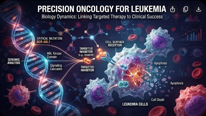
The article examines leukemia as an evolving population under targeted therapy, using chronic myeloid leukemia as the main example. BCR–ABL1 inhibition can produce deep responses, but every treatment changes the competitive environment among malignant clones. Quantitative transcript monitoring provides a continuous measure of disease burden, while deep sequencing can identify emerging resistance mechanisms before hematologic progression. A detected mutation is clinically meaningful only when its
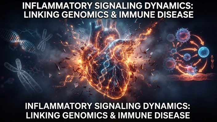
The article examines how cardiovascular genetic risk becomes biologically active through cell-specific inflammatory signaling. Because many vascular-risk variants lie in regulatory DNA rather than coding regions, their effects may appear only in particular endothelial, macrophage, smooth-muscle, or lymphocyte states. Single-cell studies of human atherosclerotic lesions and multiomic endothelial experiments are used to illustrate this context dependence. The article also discusses IL6-related sig
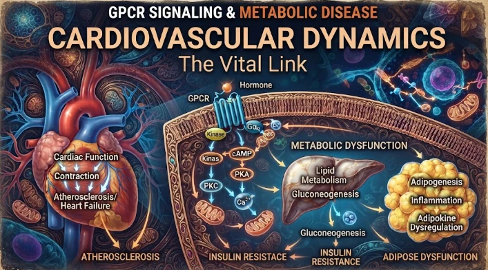
The article examines cardiovascular GPCR signaling as a dynamic response system whose abnormalities can remain hidden in resting measurements. Mechanical stretch, hormones, nutrients, sympathetic activity, and fluid balance can change the amplitude, duration, localization, and recovery of receptor signaling. Examples include mechanically activated angiotensin-receptor signaling, endosomal signaling, beta-arrestin-dependent trafficking, and biased glucagon-like peptide-one receptor responses. The