The article examines steroid receptors as dynamic transcriptional systems in hormone-dependent cancers rather than simple on–off biomarkers. Estrogen, androgen, progesterone, and glucocorticoid receptors alter gene expression through ligand-induced conformation, DNA binding, chromatin accessibility, coactivators, corepressors, and signaling crosstalk. Particular attention is given to SRC-family coactivators, enhancer architecture, growth-factor pathways, protein turnover, and mutations that change regulator stability. These mechanisms help explain why receptor-positive cancers can remain hormone dependent while becoming resistant to standard endocrine therapies. The article proposes multiomic profiling that combines receptor status with chromatin, transcriptomic, proteomic, phosphoproteomic, and single-cell information. Functional causality should then be tested through CRISPR perturbation, organoids, xenografts, and rescue experiments. Spatial and metastatic heterogeneity are also highlighted. Overall, the article argues that precision oncology should classify hormone-dependent tumors by their active regulatory mechanism and target the transcriptional machinery sustaining resistance.
The steroid receptors are at a key point in the interactions between extracellular hormones and nuclear gene control. Regulatory functions of receptor signaling in breast, prostate, endometrial and other hormone-dependent malignancies can dictate proliferation, survival, differentiation and response to therapy. Understanding the signaling as a regulatory system and not a simple switch on/off is the key for precision oncology. The transcriptional output is affected by ligand binding, receptor conformation, DNA recognition, chromatin accessibility, coactivator recruitment, and competing signaling pathways.
All members of the Nuclear receptor family are ligand regulated transcription factors. Specific DNA elements can be bound by either estrogen receptor, androgen receptor, progesterone receptor, or glucocorticoid receptor, or bound through a "tethered" interaction with other transcription factors. Binding of a hormone induces a conformational change in the receptor, such that it becomes capable of binding to a coregulator. The other partners remodel chromatin, acetylate histones, bring enhancer complexes together, and contribute to the assembly of the transcriptional machinery at target genes.
The p160 steroid receptor coactivator family illustrates how the role of the receptor is insufficient to account for tumor behavior. SRC one, two, and three enhance transcription by interacting with nuclear receptors and other signalling proteins. Many factors besides receptor gene changes can affect accumulation, phosphorylation, degradation and/or localization of the receptor that could lead to changes in the expression of hormone-dependent genes. In breast cancer, the amplification or hyperactivity in the genes of SRC 3 has specific relevance for the growth and metastatic programs.
Structural studies on ER complexes have helped decipher at molecular level how coactivator recruitment takes place. The binding of ligands induces receptor helix conformational change to the receptor surface which allows for coactivator motifs to bind. Subsequent assembly is dynamic. Different coactivators are able to be recruited in a sequential manner so as to bring about chromatin modification and transcription activation in a coordinated manner. Two ligands binding the same receptor can cause different transcription effects, which is related to the structural detail.
Enhancer architecture is another factor determining transcriptional regulation. Only a small number of steroid receptors interact with thousands of genomic locations and that number is sufficient to create a significant effect on a specific state of a cell. Lineage specific transcription factors can determine which enhancers are available, and pioneer factors can open chromatin prior to the arrival of the receptor. Receptor occupancy must then be taken into account in precision oncology in conjunction with chromatin state. A receptor positive tumor may have cells that are resistant, with a different enhancement landscape than the cells that are sensitive to treatment.
The other pathway to resistance is via signal integration. Growth factor pathways may activate receptors or coregulators to allow transcription to occur when hormone is not available. Receptor stability and coactivator function can be modified by kinases linked to the PI3K, MAPK and metabolic pathways. Study of SRC three revealed that nutrient state alters the activity of coactivators via modulation of the activity of metabolic enzymes and signaling proteins. This crosstalk is a possible reason why endocrine resistance may occur without a complete loss of the expression of receptors.
The turnover of protein is also significant. Ubiquitin mediated control can refashion transcriptional capacity as coregulators can be continually synthesized, modified and degraded. Mutations in SPOP can disrupt the degradation of proteins that regulate the activity of the androgen receptor pathway, such as SRC three, in prostate cancer. So, a change in the genome of the receptor itself can then extend the effect of the coactivator and alter the landscape of hormone-dependent transcription. Regulatory network of the receptor, not one gene, is the focus of the precision models.
There are multiple levels of this system to which therapeutic intervention can be directed. Classical endocrine therapies inhibit the production of hormones, block receptors from binding hormones, or cause degradation of receptors. Other approaches can affect the activity of coactivators, chromatin regulators, or pathways that sustain receptor-induced transcription. Three small molecules to inhibit SRC have proved that a transcriptional coregulator, which was thought to be hard to target, can become a target for drugs. This type of therapy may be combined with receptor directed therapy in certain resistant situations.
Mechanistic profiling should then go beyond the determination of mere immunohistochemistry. The abundance of receptors is clinically useful, but does not tell which enhancers are operating, which coactivators are predominant, or which kinase pathways are supporting transcription. Regulatory states under a single receptor label can be identified using chromatin accessibility assays, transcriptomics, proteomics, phosphoproteomics, and single cell profiling. Longitudinal sampling is particularly useful because of the selection and reprogramming of tumour populations by endocrine therapy.
Patient derived organoids and xenografts can test causality following molecular profiling to identify a potential resistance circuit. CRISPR perturbation can deplete a coactivator, enhancer or signaling node, and drug combinations can verify if the dependency is reversible. If disruption of a function disrupts receptor mediated transcription and re-establishes sensitivity to treatment, the molecular association is more convincing. Thereafter, such experiments connect network inference with therapeutic mechanism.
Spatial context is an additional aspect. The hormone-dependent cancers consist of tumor cells, fibroblasts, immune cells, endothelial cells and extracellular matrix areas with the potential to secrete several signaling cues. Receptors can be made more or less active by growth factors or inflammatory mediators released by neighbouring cells. Spatial transcriptomics and multiplex imaging can detect resistant niches with the presence of a reservoir of cells with different receptor signaling from the majority tumor cells. The individual niches can cause relapse even when the bulk measurements indicate effective pathway suppression.
Receptor heterogeneity from different metastatic sites must be also considered in precision oncology. The estrogen receptor expression, the abundance of coactivators or the chromatin state of a primary breast tumor and a later metastasis may be different. An evolved regulatory program may not be detected by the initial biopsy. Changes in such a manner could be detected by serial tissue sampling or by carefully validated circulating biomarkers, but interpretation in terms of function must be considered before altering therapy.
The coregulatory mechanism is layered: steroid hormone affects receptor conformation; the receptor conformation affects coregulator recruitment; chromatin state regulates accessible targets; and, signaling pathways modulate all elements. In the face of therapy, cancer cells can use any layer to continue to transcribe. Resistance can be classified by regulatory mechanism, bolstering the informative value of precision oncology.
Competition between the activating and repressing complexes is seen in the output of transcription, and corepressors must not be neglected. Certain endocrine therapeutics have partial effects that involve binding to a receptor conformation that favors corepressors rather than coactivators. If the corepressor loses its function, then the antagonist can be weak without changing the number of receptors. Resistance mechanisms not detected from studies examining only activation can be identified by profiling both sides of the regulatory balance.
This is another layer of the mechanism because of the presence of three dimensional genome organization. Steroid receptor enhancers can interact with promoters over large distances and drugs can alter these interactions. Resistant cell chromatin conformation assays can highlight the strengthening of loops and then disruption of the corresponding enhancer can be used to assess necessity. This type of architecture is one of the reasons that noncoding variants or epigenetic changes may affect the response to hormones without affecting a protein coding gene.
These mechanisms should be translated into parsimonious signatures for the development of clinical biomarkers. It will not be practical to measure each coregulator and enhancer in a routine practice. A small panel of a combination of receptor status, selected coactivator activity, pathway phosphorylation and a transcriptional response score could capture the dominant resistance class. Prospective validation would require demonstration that this classification was predictive of the benefit of a particular treatment approach and not just predictive of poor prognosis.
Receptor splice variants and altered receptor turnover can also cause drug resistance. These mechanisms can be used to retain some transcription but alter the ligand-dependence or drug-binding. These states can be identified by long read RNA sequencing and quantitative proteomics, and apparent "transcriptional escape" can be due to receptor activity or switching to another regulator, respectively, by receptor degradation assays.
A clinically useful model should incorporate multiomic at the single cell and spatial level with targeted perturbation. A design like this could help to determine which receptor associated programs grow during treatment, perform causal testing on patient models, and correlate these causal relationships to outcome. Steroid receptors are still key biomarkers but their true utility is in understanding the transcriptional machinery surrounding these receptors. Mechanistic resolution transforms hormone dependency from a blanket term into a guide to targeted therapeutic vulnerabilities.
CITE THIS ARTICLE
Tasmeem, S. (2026). Steroid Receptors Mechanisms through Transcriptional Regulation in Precision Oncology. https://www.sadiaresearchhub.com/article/c6464ab8-f0ec-5bfc-923e-87d50fde515f
S. Tasmeem, “Steroid Receptors Mechanisms through Transcriptional Regulation in Precision Oncology,” 2026. [Online]. Available: https://www.sadiaresearchhub.com/article/c6464ab8-f0ec-5bfc-923e-87d50fde515f
Tasmeem, S. (2026) ‘Steroid Receptors Mechanisms through Transcriptional Regulation in Precision Oncology’. Available at: https://www.sadiaresearchhub.com/article/c6464ab8-f0ec-5bfc-923e-87d50fde515f
Tasmeem, Sadia. “Steroid Receptors Mechanisms through Transcriptional Regulation in Precision Oncology.” 2026, https://www.sadiaresearchhub.com/article/c6464ab8-f0ec-5bfc-923e-87d50fde515f.
Sadia Tasmeem. “Steroid Receptors Mechanisms through Transcriptional Regulation in Precision Oncology.” (2026). https://www.sadiaresearchhub.com/article/c6464ab8-f0ec-5bfc-923e-87d50fde515f.
Tasmeem S. Steroid Receptors Mechanisms through Transcriptional Regulation in Precision Oncology. 2026. https://www.sadiaresearchhub.com/article/c6464ab8-f0ec-5bfc-923e-87d50fde515f
@misc{article_c6464ab8f0ec5bfc923e87d50fde515f,
  title = {Steroid Receptors Mechanisms through Transcriptional Regulation in Precision Oncology},
  author = {Sadia Tasmeem},
  year = {2026},
  url = {https://www.sadiaresearchhub.com/article/c6464ab8-f0ec-5bfc-923e-87d50fde515f},
}
TY  - GEN
AU  - Sadia Tasmeem
TI  - Steroid Receptors Mechanisms through Transcriptional Regulation in Precision Oncology
PY  - 2026-07-05
UR  - https://www.sadiaresearchhub.com/article/c6464ab8-f0ec-5bfc-923e-87d50fde515f
ER  -
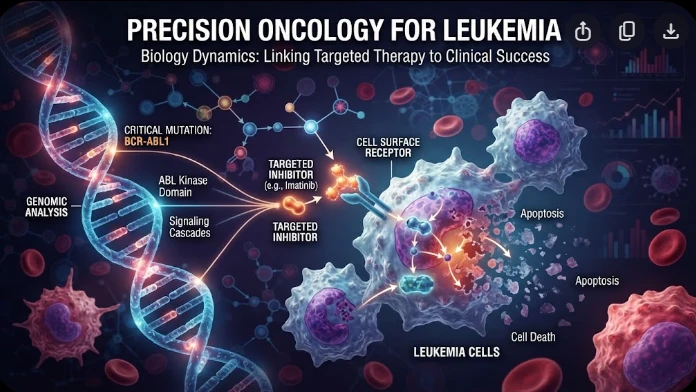
The article examines leukemia as an evolving population under targeted therapy, using chronic myeloid leukemia as the main example. BCR–ABL1 inhibition can produce deep responses, but every treatment changes the competitive environment among malignant clones. Quantitative transcript monitoring provides a continuous measure of disease burden, while deep sequencing can identify emerging resistance mechanisms before hematologic progression. A detected mutation is clinically meaningful only when its
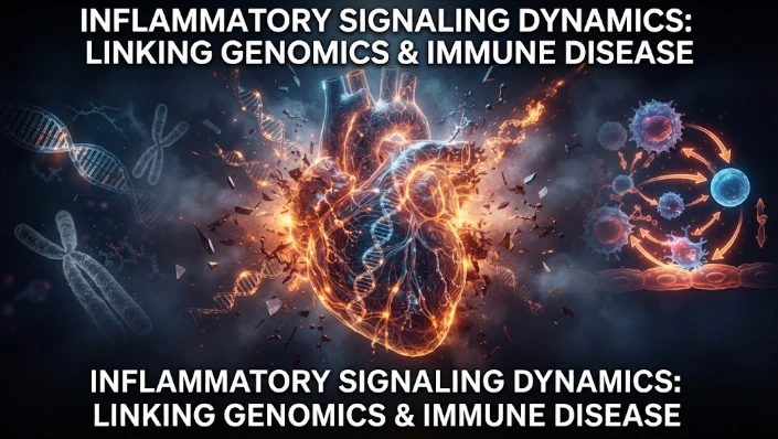
The article examines how cardiovascular genetic risk becomes biologically active through cell-specific inflammatory signaling. Because many vascular-risk variants lie in regulatory DNA rather than coding regions, their effects may appear only in particular endothelial, macrophage, smooth-muscle, or lymphocyte states. Single-cell studies of human atherosclerotic lesions and multiomic endothelial experiments are used to illustrate this context dependence. The article also discusses IL6-related sig
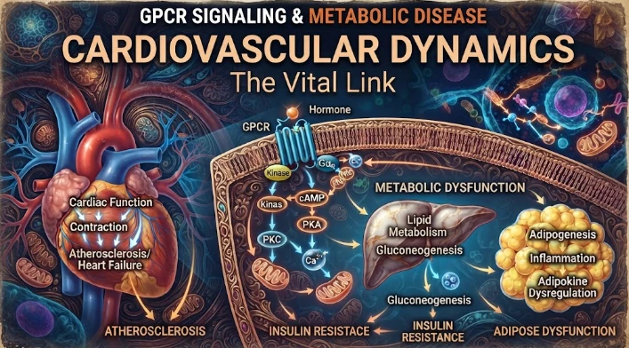
The article examines cardiovascular GPCR signaling as a dynamic response system whose abnormalities can remain hidden in resting measurements. Mechanical stretch, hormones, nutrients, sympathetic activity, and fluid balance can change the amplitude, duration, localization, and recovery of receptor signaling. Examples include mechanically activated angiotensin-receptor signaling, endosomal signaling, beta-arrestin-dependent trafficking, and biased glucagon-like peptide-one receptor responses. The